
Page 555 - Feline diagnostic imaging
P. 555
(b)
(a)
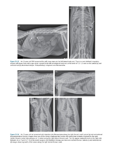
Figure 31.15 An 11-year-old DSH presented for soft tissue mass on the left lateral body wall. There is a well-defined, irregularly
shaped soft tissue mass with a gas center located in the left dorsolateral body wall at the level of T13–L3 seen on the lateral (a) and
ventrodorsal (b) abdominal images. Histopathologic diagnosis was fibrosarcoma.
(a) (c)
(b) (d)
Figure 31.16 An 11-year-old cat presented with injection site fibrosarcoma along the right thoracic wall. Lateral (a) and ventrodorsal
(b) postoperative thoracic images show loss of the normal diaphragmatic border with abdominal contents displaced to the right
caudal thoracic cavity. Subcutaneous air is noted along the right lateral thoracic wall. The ribs appear malpositioned on the right side
between rib 6–7, and rib 7–8 at the recent surgical site. Radiation therapy was done and recheck thoracic lateral (c) and ventrodorsal
(d) images show regrowth of the tumor along the right lateral thoracic wall.