
Page 559 - Feline diagnostic imaging
P. 559
572 31 Body Wall
(a) (b)
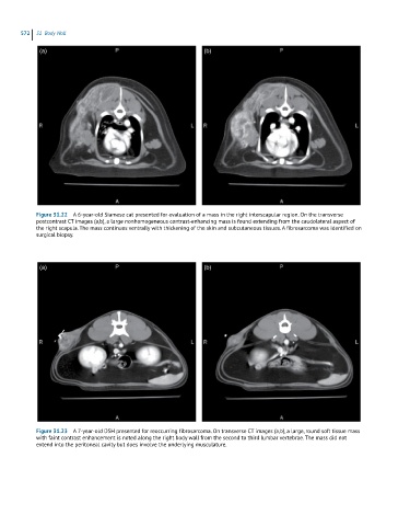
Figure 31.22 A 6-year-old Siamese cat presented for evaluation of a mass in the right interscapular region. On the transverse
postcontrast CT images (a,b), a large nonhomogeneous contrast-enhancing mass is found extending from the caudolateral aspect of
the right scapula. The mass continues ventrally with thickening of the skin and subcutaneous tissues. A fibrosarcoma was identified on
surgical biopsy.
(a) (b)
Figure 31.23 A 7-year-old DSH presented for reoccurring fibrosarcoma. On transverse CT images (a,b), a large, round soft tissue mass
with faint contrast enhancement is noted along the right body wall from the second to third lumbar vertebrae. The mass did not
extend into the peritoneal cavity but does involve the underlying musculature.