
Page 558 - Feline diagnostic imaging
P. 558
31.3 SAc Diiss sSopeiDe SAects Sody epp 571
(a) (b)
(c)
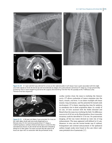
Figure 31.20 A 7-year-old DSH was referred for a mass on the right shoulder. A soft tissue mass is seen associated with the right
proximal scapula on both the lateral (a) and ventrodorsal (b) images. On a postcontrast transverse CT image (c), a large peripherally
enhancing mass is found beginning lateral to the scapula and extending from the first rib caudally to the midthoracic area.
Fibrosarcoma was diagnosed.
cardiac motion when the mass is overlying the thoracic
cavity. CT allows evaluation of tumor extension into the
fascia, muscle, and bone. It can detect multiple soft tissue
masses, lung metastasis, and the potential for lymph node
involvement. CT is faster, requiring less time for sedation
or anesthesia due to short acquisition times. In a study of
22 cats, 10 were scanned with the limbs extended and
flexed when the mass was overlying the limb. No difference
was seen in 8/10 but on the flexed position, fewer muscular
invasions could be identified in 2/10 cats. On postcontrast
Figure 31.21 A 14-year-old Maine Coon presented for mass on imaging, all but one tumor showed an outer rim of ring
the right lateral body wall previously diagnosed as a enhancement. The mass appeared well defined in 13 cats.
fibrosarcoma. On the transverse postcontrast CT image, a However, in nine cats the ventral border was ill defined.
nonhomogeneous contrast enhancement mass is present in the Enlargement and contrast enhancement of the ipsilateral
subcutaneous tissues on the dorsal lateral abdomen extending
along the lumbar spine. The mass extends into the superficial axillary lymph nodes were found in five cats which indi-
muscular layer with no extension into the peritoneal cavity. cates potential site of metastasis [17].