
Page 560 - Feline diagnostic imaging
P. 560
31.3 SAc Diiss sSopeiDe SAects Sody epp 573
(a) (b)
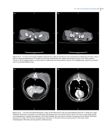
Figure 31.24 A 5-year-old domestic longhair presented for a mass on the left rear limb. A peripherally enhancing soft tissue mass is
seen on transverse postcontrast CT images (a,b); the mass extends from the lateral subcutaneous tissues to the vastus lateralis
muscle. In other imaging planes this mass may be invading into the biceps femoris muscle. The histopathologic diagnosis was feline
vaccine-associated fibrosarcoma.
(a) (b)
Figure 31.25 A 14-year-old DSH presented for a mass on the left thoracic wall. On a postcontrast transverse CT image (a), a large
ill-defined ovoid mass is present on the left dorsal subcutaneous tissue along the thoracic cavity. The mass is a soft tissue opacity
with heterogeneous contrast enhancement. The borders between the mass and the epaxial musculature are ill defined. Transverse
postcontrast image of the thoracic cavity (b) shows thickening of the left ventricular wall consistent with hypertrophic
cardiomyopathy. The mass was diagnosed as a fibrosarcoma.