
Page 557 - Feline diagnostic imaging
P. 557
570 31 Body Wall
to find on radiographs alone. CT postcontrast images planning even after surgical excision to determine the radi-
typically show the tumor to be twice the size appreciated ation treatment area [16].
by palpation alone. Alternatively, due to its superior soft A more recent review article of feline injection site sar-
tissue imaging, MRI can be used to determine the extent coma (FISS) found a higher incidence of tumor with
of disease. CT with contrast has been used for treatment increased numbers of vaccinations per site, injection into
the interscapular region, and if vaccines were administered
cold versus at room temperature. Staging of FISS for
metastasis to the lungs occurs in 10–25% of cases and should
be evaluated by three-view thoracic imaging or thoracic CT.
The regional lymph nodes should be palpated and sampled
by cytologic evaluation. Additional sites of metastasis
include the liver and subcutaneous tissue. MRI or CT may
overestimate the size of the tumor but this may improve
complete surgical resection of the mass. It is unclear if
advanced imaging improves survival times but it does assist
surgical planning. Recurrence at the surgical site even with
clean margins occurs in 14–50% of sites. It is speculated that
surgical manipulation of the tissue may incite recurrence of
the tumor. Radiotherapy performed before or after surgical
excision has been advocated, although the benefit has not
been conclusively determined [15].
31.3.1.1 Computed Tomography of FISS
The typical location of FISS within the soft tissues is over
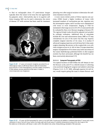
Figure 31.18 A 7-year-old domestic longhair presented for an the thoracic cavity and CT is a reliable means of collecting
interscapular mass. A soft tissue mass is seen on transverse
postcontrast CT with heterogeneous contrast uptake. Irregularity cross-sectional imaging for surgical and radiation planning
of the spinous process adjacent to the mass is noted. A (Figures 31.21–31.27). MRI, although superior for soft tis-
chondrosarcoma was diagnosed. sue, would require gating the exam to lessen thoracic and
(a) (b)
Figure 31.19 A 9-year-old DSH presented for mass on the left flank. A postsurgical radiation treatment planning CT study (a,b) shows
extension of abnormal soft tissue within the body wall with focal areas of contrast enhancement adjacent to the surgical site.
No extension of abnormal soft tissue is seen within the abdominal cavity. Histopathology revealed a fibrosarcoma.