Page 556 - Feline diagnostic imaging
P. 556
31.3 SAc Diiss sSopeiDe SAects Sody epp 569
(a) (b)
(c)
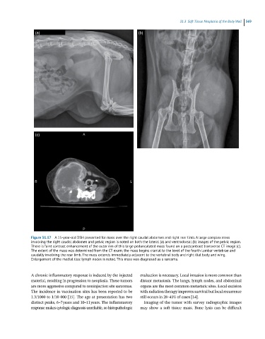
Figure 31.17 A 15-year-old DSH presented for mass over the right caudal abdomen and right rear limb. A large complex mass
involving the right caudal abdomen and pelvic region is noted on both the lateral (a) and ventrodorsal (b) images of the pelvic region.
There is faint contrast enhancement of the outer rim of this large pedunculated mass found on a postcontrast transverse CT image (c).
The extent of the mass was determined from the CT exam; the mass begins cranial to the level of the fourth lumbar vertebrae and
caudally involving the rear limb. The mass extends immediately adjacent to the vertebral body and right ilial body and wing.
Enlargement of the medial iliac lymph nodes is noted. This mass was diagnosed as a sarcoma.
A chronic inflammatory response is induced by the injected evaluation is necessary. Local invasion is more common than
material, resulting in progression to neoplasia. These tumors distant metastasis. The lungs, lymph nodes, and abdominal
are more aggressive compared to noninjection site sarcomas. organs are the most common metastatic sites. Local excision
The incidence in vaccination sites has been reported to be with radiation therapy improves survival but local recurrence
1.3/1000 to 1/10 000 [15]. The age at presentation has two still occurs in 28–45% of cases [14].
distinct peaks, 6–7 years and 10–11 years. The inflammatory Imaging of the tumor with survey radiographic images
response makes cytologic diagnosis unreliable, so histopathologic may show a soft tissue mass. Bone lysis can be difficult

