
Page 552 - Feline diagnostic imaging
P. 552
(b)
(a)
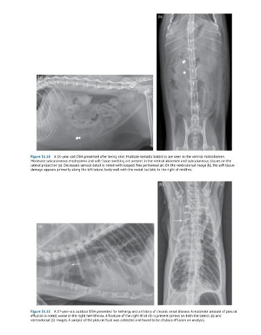
Figure 31.10 A 10-year-old DSH presented after being shot. Multiple metallic ballistics are seen in the ventral midabdomen.
Moderate subcutaneous emphysema and soft tissue swelling are present in the ventral abdomen and subcutaneous tissues on the
lateral projection (a). Decreased serosal detail is noted with suspect free peritoneal air. On the ventrodorsal image (b), the soft tissue
damage appears primarily along the left lateral body wall with the metal ballistic to the right of midline.
(b)
(a)
Figure 31.11 A 17-year-old outdoor DSH presented for lethargy and a history of chronic renal disease. A moderate amount of pleural
effusion is noted, worse in the right hemithorax. A fracture of the right third rib is present (arrow) on both the lateral (a) and
ventrodorsal (b) images. A sample of the pleural fluid was collected and found to be chylous effusion on analysis.