
Page 548 - Feline diagnostic imaging
P. 548
31.2 Diseisi AAsecDing ctse Dii 561
(b)
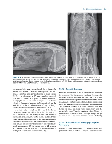
(a)
Figure 31.4 A 6-year-old DSH presented for dysuria of one day’s duration. There is mottling of the subcutaneous tissues along the
ventral abdominal wall on the lateral image (a). On the ventrodorsal image (b), there is mild asymmetry and increase in the external
soft tissues adjacent to the right caudal abdominal wall compared to the left. This should not be confused with acute trauma and may
be related to recent subcutaneous fluid administration.
contrast resolution and improved resolution of tissues of a 31.2.8 Magnetic Resonance
similar density make CT superior to radiographs. Improved Magnetic resonance (MR) has superior contrast resolution
spatial resolution enables visualization of small lesions
0.2–0.3 mm in diameter. As CT technology has improved, for soft tissue. Use in veterinary medicine for superficial
neoplasia is slowly increasing but continues to be limited
less time is required to acquire the images. Computed
tomographic studies are used in surgical and radiation due to cost and need for general anesthesia. In human medi-
cine, dynamic contrast-enhanced magnetic resonance imag-
planning. Contrast enhancement of tumors involving the
soft tissue and bone and evaluation of regional lymph ing (MRI) studies evaluate the contrast perfusion of a tumor.
The contrast is followed as it enters, enhances, and then
nodes for metastasis can be documented with CT [9].
In a study using whole-body CT to check for distant leaves the tumor assessing vessel permeability and the
metastases, masses were identified within the muscle in extracellular space. This aids in determination of benign or
6/193 cats examined. The primary tumors were located in normal tissue versus malignant, although histopathologic
evidence of tumor can persist even with a normal study [11].
the mammary glands, oral cavity, and mediastinal lymph
node. The pathologic diagnosis of the muscle masses was
carcinoma in five cases and lymphoma in one. On precon-
trast images, the masses were isodense to the normal mus- 31.2.9 Positron Emission Tomography/Computed
Tomography
cle. On postcontrast CT, the masses were ovoid to round
with varying degrees of contrast enhancement helping to Positron emission tomography (PET) scans are commonly
distinguish borders from normal tissues [10]. performed in human medicine using a radiopharmaceutical