
Page 561 - Feline diagnostic imaging
P. 561
574 31 Body Wall
vascularity in the outer portions of the tumor, with no per-
fusion detected in the center [19].
31.3.2 Mammary Gland Neoplasia
Mammary gland neoplasia is the third most common neo-
plasia in the cat (Figures 31.28 and 31.29). These tumors
are hormonally driven with a higher incidence in female
cats spayed after 1 year of age. Malignant neoplasia is more
common than benign, with most common being adenocar-
cinoma (carcinoma). Some studies suggest the caudal
mammary glands are predisposed to neoplasia. Benign
neoplasia accounts for 10–20% of mammary masses, with
fibroadenoma the most frequently reported. Other
nonneoplastic mammary gland changes are associated
with fibroadenomatous hyperplasia due to hormonal
response or treatment with progestational compounds.
Fibroadenomas typically occur in young female cats pre-
senting with one or multiple enlarged mammary glands,
some becoming edematous and ulcerated. The prognosis
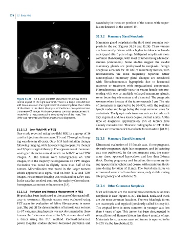
Figure 31.26 An 8-year-old DSH presented for a mass on the
lateral aspect of the right rear limb. There is a large, well-defined worsens when the size of the tumor exceeds 3 cm. The rate
soft tissue mass on the right hindlimb extending from the middle of metastasis is reported to be 50–90%, with the regional
of the ilium to the distal diaphysis of the femur on a postcontrast lymph nodes and lungs being the most common sites for
transverse CT image. Nonhomogeneous contrast enhancement is metastasis. The lymph node involvement can include axil-
noted with a hypoattenuating central region of the mass. The
limb was removed and fibrosarcoma was diagnosed. lary, inguinal, and, to a lesser degree, sternal nodes. At the
time of diagnosis, approximately 25% of tumors have
already metastasized. Thoracic radiographs or CT of the
31.3.1.2 Low-Field MRI of FISS thorax are recommended to evaluate for metastasis [20,21].
One study reported using low-field MRI in a group of 19
cats for injection site sarcomas. T1- and T2-weighted imag- 31.3.3 Mammary Gland Ultrasound
ing was done in all cases. Only 5/19 had radiation therapy
following imaging, with 3/5 receiving preoperative therapy Ultrasound evaluation of 35 female cats, 13 nonpregnant,
and 2/5 postsurgical therapy. The appearance of the tumor six early pregnancy, eight late pregnancy, and 16 lactating
was hyperintense to normal muscle on both T1W and T2W cats was performed. In the nonpregnant cats, the mam-
images. All the tumors were heterogeneous on T2W mary tissue appeared hypoechoic and less than 2.0 mm
images, with the majority heterogeneous on T1W images. thick. During pregnancy and lactation, the mammary tis-
Cavitation was noted in slightly more than half of the sue appears hyperechoic and coarse, with maximum thick-
tumors. Mineralization was noted in the larger tumors ness during lactation of 11 mm. The ductal structures on
which appeared as a signal void on both T1W and T2W ultrasound were small anechoic areas, only visible during
images. Postcontrast imaging was evaluated in 12/19 cats. late pregnancy and lactation [22].
In the cats that received contrast, the majority showed non-
homogeneous contrast enhancement [18]. 31.3.4 Other Cutaneous Neoplasia
31.3.1.3 Perfusion and Hypoxia Measurement in FISS Mast cell tumors are the second most common cutaneous
Hypoxia has been implicated as the cause of decreased effi- neoplasia in cats (Figure 31.30). The head, neck, and truck
cacy to treatment. Hypoxia tracers were evaluated using are the most common locations. The two histologic forms
PET scans for evaluation of feline fibrosarcoma in seven are mastocytic and atypical (previously called histiocytic).
cats. The cut-off for determination of hypoxia was not met The atypical form is more common in younger cats less
in 4/7 cats, meaning hypoxia was not documented in these than 4 years of age. This tumor has been documented in
tumors. Perfusion was elevated in 5/7 cats examined with several litters of Siamese kittens less than 6 months of age.
a tracer using the PET method. Contrast-enhanced Metastasis for cutaneous mast cell tumor is reported to be
power Doppler studies showed decreased perfusion and 0–22% via the lymphatics [23].