
Page 534 - Feline diagnostic imaging
P. 534
546 30 Peritoneal Cavity
hernia. Loss of normal diaphragmatic contour, displace- cats with a lower respiratory rate on presentation had a
ment of a liver lobe, or positive contrast leakage into the higher mortality rate [15].
thoracic cavity are consistent with a diaphragmatic hernia. At least two radiographic tangential projections are
Chronic diaphragmatic hernia with the presence of pleural needed for accurate diagnosis of a diaphragmatic hernia
or peritoneal effusion will be more difficult to identify with (Figures 30.25 and 30.26). Right‐sided hernias are more fre-
this technique since contrast may not flow due to fibrosis quently found. Reported radiographic changes associated
and adhesions at the herniation site. with diaphragmatic hernia are alterations in the diaphrag-
Celiography is contraindicated in patients with peritoni- matic shape, position, or loss of visualization of the dia-
tis, hypovolemia, impaired renal function, or known phragmatic contour. Within the thoracic cavity, an increase
adverse reactions to contrast agents [14]. in soft tissue was the most consistent finding with a dia-
phragmatic hernia. In addition, varying degrees of pleural
30.5.3 Diaphragmatic Hernia effusion, mediastinal shift, cardiac displacement, pneumo-
thorax, or dorsal tracheal displacement can be observed.
Congenital diaphragmatic hernias are rare in cats. The lung parenchyma displacement and compression will
Traumatically induced diaphragmatic hernias are usu- aid in identification of the diaphragmatic hernia site. In
ally secondary to blunt force trauma, with the most most cases, the abdominal organs are displaced cranially
reported reason being vehicular trauma. The most com- with the stomach or small intestinal loops within the tho-
mon clinical sign on presentation was tachypnea. The racic cavity. Concurrent rib fractures are seen in a small
liver was the most frequently herniated organ in a group number of cats [16].
of 34 cats with diaphragmatic hernias. The duration of In a study of 16 cats with chronic diaphragmatic hernia,
the hernia did not affect the outcome. However, older defined as herniation present longer than two weeks, the
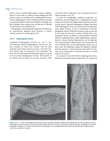
(b)
(a)
Figure 30.25 A 2-year-old female DSH presented due to increased respiratory effort and muffled heart sounds. (a) A lateral thoracic
radiograph shows the majority of the abdominal contents within the thoracic cavity. (b) Ventrodorsal thoracic radiograph. The cardiac
silhouette is shifted to the right of the midline. The diaphragm is not visualized from the midline to the left side. A diaphragmatic
hernia was corrected at surgery.