Page 568 - Feline diagnostic imaging
P. 568
582  32  Overview of the Musculoskeletal System


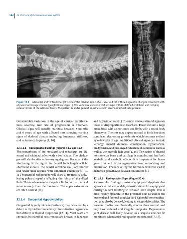
            Figure 32.1  Lateral (a) and ventrodorsal (b) views of the cervical spine of a 1-year-old cat with radiographic changes consistent with
            a lysosomal storage disease (gangliosidosis type II). The vertebrae are abnormal in shape with ill-defined endplates and bridging
            osteoarthrosis of the articular facets. The patient is under general anesthesia with an endotracheal tube present.


            Considerable variation in the age of clinical manifesta-  and Abyssinian cats [1]. The most obvious clinical signs are
            tion,  severity,  and  rate  of  progression  is  observed.   those of disproportionate dwarfism. These include a large
            Clinical signs will usually manifest between 5 months   broad head with a short neck and limbs with a round body
            and 6 years of age with affected cats showing varying   phenotype. The cats may appear normal at birth but show
            signs  of  skeletal  disease  including  lameness,  stiffness,   significant decreasing growth rate which becomes evident
            and reluctance to jump [1, 10].                   by 6–8 weeks of age. Additional clinical signs can include
                                                              lethargy,  mental  dullness,  constipation,  hypothermia,
            32.1.3.1  Radiographic Findings (Figures 32.2 and 32.3)  bradycardia, and prolonged retention of deciduous teeth as
            The  metaphyses  of  the  metatarsi  and  metacarpi  are  dis-  well as the juvenile hair coat [1, 15]. The action of thyroid
            torted and widened, often with a bent shape. The phalan-  hormone on bone and cartilage is complex and has both
            ges will also be affected to varying degrees. Because of the   anabolic  and  catabolic  effects.  It  is  important  for  linear
            shortening  of  the  digits,  the  overall  limb  length  will  be   growth  as  well  as  for  appropriate  bone  remodeling  and
            shortened as well. The caudal vertebrae (tail) are shorter   maturation. The lack of thyroid hormone will thus lead to
            and  wider  than  normal  with  abnormal  endplates  [7,  10,   disturbed growth and delayed maturation [1].
            11]. Sequential radiographs will show a progressive anky-
            losing  polyarthropathy  affecting  the  joints  of  the  distal   32.1.4.1  Radiographic Signs (Figure 32.4)
            limb. This tends to involve the pelvic limbs both earlier and   Radiographic findings consist of epiphyseal dysplasia that
            more  severely  than  the  forelimbs. The  upper  extremities   appears as reduced or delayed ossification of the epiphyseal
            are often normal [10].                            cartilage  model  resulting  in  reduced  limb  length. This  is
                                                              most readily apparent in the proximal tibia as well as the
                                                              humeral and femoral condyles [15]. Cuboidal bone ossifica-
            32.1.4  Congenital Hypothyroidism
                                                              tion may also be delayed, leading to valgus deformities. The
            Congenital hypothyroidism (cretinism) may be caused by a   vertebral  bodies  are  classically  shorter  than  normal  and
            defect in thyroid hormone biosynthesis (iodine organifica-  may  have  widened  and  irregular  endplates.  Degenerative
            tion defect) or thyroid dysgenesis [12–14]. Most cases are   joint  disease  will  likely  develop  as  a  sequela  and  can  be
            sporadic, but familial occurrences are known in Japanese   monitored when serial radiographs are obtained [7, 15].

