
Page 569 - Feline diagnostic imaging
P. 569
32.1 uvenile one Disease 583
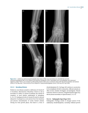
Figure 32.2 Lateral (a,c) and dorsopalmar/plantar (b,d) views of the carpus and tarsus in a Scottish fold cat with
osteochondrodysplasia. Notice the widened and distorted metaphysis of the metacarpal bones and phalanges with shortened
appearing digits. There is also ankylosing arthropathy present in the carpus and tarsus along with new bone production at insertion
points of the calcaneus. These are all common features of osteochondrodysplasia. Source: Courtesy of Dr Jay Griffin.
32.1.5 Hereditary Rickets chondrodysplasia [1]. Cartilage will continue to accumulate
in the metaphysis, but fail to mineralize. Affected animals are
Rickets is a rare disease caused by a deficiency of vitamin D often severely stunted, bowlegged, and plantigrade. Muscle
or hereditary abnormalities. Hereditary rickets in cats is weakness, tremors or seizures, and gastrointestinal signs may
secondary to a defect in vitamin D pathway, the vitamin D also be present secondary to hypocalcemia [1, 16, 17].
receptors, or renal tubular reabsorption of phosphate
[1, 16, 17]. Inadequate levels of vitamin D result in reduced
absorption of phosphorus and calcium from the intestines 32.1.5.1 Radiographic Signs (Figure 32.5)
and prevent mineralization of newly formed osteoid. Radiographic findings consist of changes typical of the
During the bone growth phase, this leads to a form of underlying chondrodysplasia, including widened growth