
Page 570 - Feline diagnostic imaging
P. 570
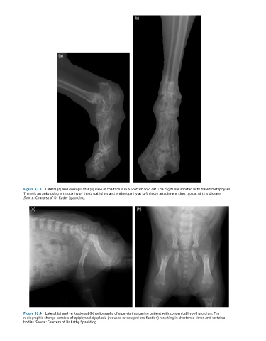
Figure 32.3  Lateral (a) and dorsoplantar (b) view of the tarsus in a Scottish fold cat. The digits are shorted with flared metaphyses.
            There is an ankylosing arthropathy of the tarsal joints and enthesopathy at soft tissue attachment sites typical of this disease.
            Source: Courtesy of Dr Kathy Spaulding.


            Figure 32.4  Lateral (a) and ventrodorsal (b) radiographs of a pelvis in a canine patient with congenital hypothyroidism. The
            radiographic change consists of epiphyseal dysplasia (reduced or delayed ossification) resulting in shortened limbs and vertebral
            bodies. Source: Courtesy of Dr Kathy Spaulding.