
Page 575 - Feline diagnostic imaging
P. 575
32.2 Accuired one Disease 589
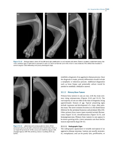
Figure 32.10 Oblique lateral views of the left and right antebrachii in an 8-month-old kitten. There is bilateral radial hemimelia, the
most common type of hemimelia reported in cats. It is often unilateral but in this kitten it was bilateral. This defect has resulted in
severe angular limb deformity and poorly developed carpi.
establish a diagnosis of an aggressive disease process. Once
the diagnosis is made, primary differentials would include
a neoplastic or infectious process. Additional diagnostics
such as bone biopsy and potentially culture would be
needed to establish a definitive answer.
32.2.2 Primary Bone Tumors
Primary bone tumors in cats are rare, with the most com-
mon being osteosarcoma (Figures 32.12 and 32.13). The
vast majority of cats are older at the time of diagnosis, being
approximately 10 years of age. Typical presenting signs
include lameness and development of a large, often pain-
less mass. The most common location is in the distal femur
followed by the proximal humerus and proximal tibia [30].
Other primary bone tumor types reported include fibrosar-
coma (Figure 32.14), chondrosarcoma (Figure 32.15), and
hemangiosarcoma. Primary bone tumors in cats appear to
be slower growing with a lower metastatic rate than similar
tumors reported for dogs [30–32].
Figure 32.11 Lateral (a) and craniocaudal (b) views of the 32.2.2.1 Radiographic Signs
antebrachium of a cat with unilateral radial hemimelia. There is The radiographic appearance is variable and typical of an
corresponding luxation of the carpus and marked angular limb
changes typical with this anomaly. Source: Courtesy of Dr Jay aggressive disease response. Lesions are usually monosto-
Griffin. tic, metaphyseal, and can be purely lytic, proliferative, or