
Page 579 - Feline diagnostic imaging
P. 579
32.2 Accuired one Disease 593
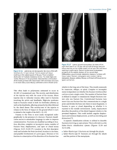
Figure 32.17 Lateral (a) and dorsopalmar (b) views of the
right front foot in an 18-year-old cat with lameness and soft
tissue swelling on the medial aspect of the paw. The soft tissue
Figure 32.16 Lateral (a) and dorsoplantar (b) views of the left swelling is asymmetric. There is subtle bone involvement
hind foot of a 12-year-old cat. There is severe soft tissue primarily of the middle phalanx of the second digit.
swelling on the lateral aspect of the pes. There is marked Differentials would include metastatic disease or primary soft
destruction of multiple metatarsal bones. The distal digits are tissue tumor. Thoracic radiographs were normal, making
normal. Because of the polyostotic characteristics and location metastatic disease unlikely. Biopsy confirmed soft tissue tumor
of the lesion, primary soft tissue tumor with secondary bony lysis (giant cell tumor).
is the most likely cause. Biopsy confirmed a nerve sheath tumor.
relative to the long axis of the bone. This would commonly
be transverse, oblique, or spiral. Complete or incomplete
This often leads to polytrauma estimated to occur in status is dependent on whether the fracture involves either
30–60% of traumatized cats. The severity and distribution cortices or just a single cortex. The number of fracture lines
of the injuries vary with the cause of the trauma. Blunt is defined as simple or comminuted. Simple fractures con-
injuries due to vehicular trauma often lead to fractures tain only one fracture line. Comminuted fractures have
involving the pelvis and hindlimbs. High-rise syndrome more than one fracture line that communicates to a single
leads to fractures noted in both the forelimbs (elbows) as point and divides the bone into three or more fragments. A
well as the hindlimbs, affecting primarily the tibia followed fracture is open or closed depending on whether it is
by the distal femur. The severity/rate of the injuries is exposed to the outside environment. Lastly, displacement
related to the force of impact on the ground which in turn describes the position of the distal fragment relative to the
is related to the height of the fall [1, 42, 43]. proximal fragment. This could include angular displace-
Trauma to the bone is most easily recognized radio-
graphically by the presence of a fracture. Fracture classifi- ment and torsional displacement, as well as overriding and
distraction [7].
cation serves to standardize language in order to improve A separate classification scheme is utilized to describe
communication. Fractures are classified according to loca- fractures involving an open physis. This is referred to as the
tion, direction, complete or incomplete status, number of Salter–Harris classification system (Figures 32.30 and
fracture lines, displacement, and open or closed status 32.31) [44].
(Figures 32.23–32.29) [7]. Location is the first descriptor
used and includes the bone involved, location in the bone, ● Salter–Harris type I: fractures are through the physis.
and possible involvement of a joint space. Direction of the ● Salter–Harris type II: fractures are through the physis
fracture is a description of the direction of the fracture line and the portion of the metaphysis.