
Page 580 - Feline diagnostic imaging
P. 580
594 32 Overview of the Musculoskeletal System
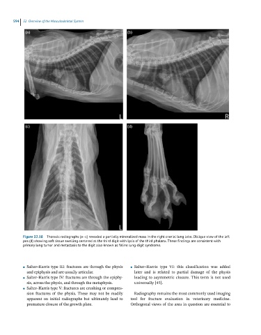
Figure 32.18 Thoracic radiographs (a–c) revealed a partially mineralized mass in the right cranial lung lobe. Oblique view of the left
pes (d) showing soft tissue swelling centered at the third digit with lysis of the third phalanx. These findings are consistent with
primary lung tumor and metastasis to the digit also known as feline lung-digit syndrome.
Salter–Harris type III: fractures are through the physis Salter–Harris type VI: this classification was added
● ●
and epiphysis and are usually articular. later and is related to partial damage of the physis
Salter–Harris type IV: fractures are through the epiphy- leading to asymmetric closure. This term is not used
●
sis, across the physis, and through the metaphysis. universally [45].
Salter–Harris type V: fractures are crushing or compres-
●
sion fractures of the physis. These may not be readily Radiography remains the most commonly used imaging
apparent on initial radiographs but ultimately lead to tool for fracture evaluation in veterinary medicine.
premature closure of the growth plate. Orthogonal views of the area in question are essential to