
Page 582 - Feline diagnostic imaging
P. 582
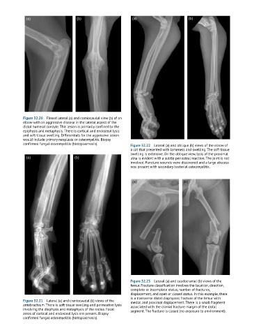
Figure 32.20  Flexed lateral (a) and craniocaudal view (b) of an
            elbow with an aggressive disease in the lateral aspect of the
            distal humeral condyle. This lesion is primarily confined to the
            epiphysis and metaphysis. There is cortical and endosteal lysis
            and soft tissue swelling. Differentials for the aggressive lesion
            would include primary neoplasia or osteomyelitis. Biopsy
            confirmed fungal osteomyelitis (histoplasmosis).  Figure 32.22  Lateral (a) and oblique (b) views of the elbow of
                                                              a cat that presented with lameness and swelling. The soft tissue
                                                              swelling is extensive. On the oblique view, lysis of the proximal
                                                              ulna is evident with a subtle periosteal reaction. The joint is not
                                                              involved. Puncture wounds were discovered and a large abscess
                                                              was present with secondary bacterial osteomyelitis.


                                                              Figure 32.23  Lateral (a) and caudocranial (b) views of the
                                                              femur. Fracture classification involves the location, direction,
                                                              complete or incomplete status, number of fractures,
                                                              displacement, and open or closed status. In this example, there
                                                              is a transverse distal diaphyseal fracture of the femur with
            Figure 32.21  Lateral (a) and craniocaudal (b) views of the   medial and proximal displacement. There is a small fragment
            antebrachium. There is soft tissue swelling and permeative lysis   associated with the cranial fracture margin of the distal
            involving the diaphysis and metaphysis of the radius. Focal   segment. The fracture is closed (no exposure to environment).
            areas of cortical and endosteal lysis are present. Biopsy
            confirmed fungal osteomyelitis (histoplasmosis).