
Page 587 - Feline diagnostic imaging
P. 587
32.3 oint Disease 601
Figure 32.35 Lateral radiographs of a tibia of a juvenile feline
patient with a distal diaphyseal spiral fracture of the tibia (a).
The fibula was intact. Conservative treatment was instituted.
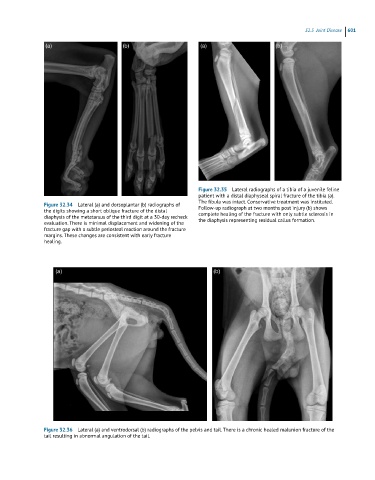
Figure 32.34 Lateral (a) and dorsoplantar (b) radiographs of Follow-up radiograph at two months post injury (b) shows
the digits showing a short oblique fracture of the distal complete healing of the fracture with only subtle sclerosis in
diaphysis of the metatarsus of the third digit at a 30-day recheck the diaphysis representing residual callus formation.
evaluation. There is minimal displacement and widening of the
fracture gap with a subtle periosteal reaction around the fracture
margins. These changes are consistent with early fracture
healing.
Figure 32.36 Lateral (a) and ventrodorsal (b) radiographs of the pelvis and tail. There is a chronic healed malunion fracture of the
tail resulting in abnormal angulation of the tail.