
Page 588 - Feline diagnostic imaging
P. 588
602 32 Overview of the Musculoskeletal System
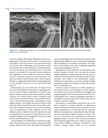
Figure 32.37 Osteopetrosis diagnosed in a cat. Lateral (a) and ventrodorsal (b) images of the pelvis showed increased variable
opacity in all bony structures.
articular cartilage. With benign disease processes such as such as cranial displacement of the tibia with cranial cruciate
degenerative joint disease that results in increased stress ligament injury (Figures 32.43 and 32.44), elbow incongruity,
remodeling of the joint surfaces, the subchondral bone and hip dysplasia are less obvious. Nonetheless, joint align-
will become more radiopaque and potentially thicker ment is an important factor when considering underlying
than normal. This is often referred to as subchondral scle- causes of joint pathology [7, 54, 55].
rosis and will appear as a subchondral zone of increased Osteophytes are bony outgrowths at the periphery of the
opacity 1–2 mm wide [7]. Conversely, if the joint disease is articular margins of joints. They are often the primary
more aggressive, such as would occur with severe inflam- change identified in osteoarthrosis and are the result of
matory or infectious causes, the subchondral bone may ossification of chondrocytes formed from pluripotent mes-
show overt signs of lysis and destruction. This will appear enchymal cells located at the osteochondral margins.
initially as a ragged subchondral bone margin progressing Although the mechanisms are unclear, articular cartilage
to near-complete destruction of the subchondral bone degradation products are thought to mediate the develop-
plate [7]. ment of osteophytes [56].
Mineralization can occur within the soft tissues of the Enthesis is a point of insertion of a tendon, ligament, or
joint (synovial membrane, joint capsule, supporting struc- joint capsule to bone. An enthesophyte is a bony prolifera-
tures) or within the joint compartment as a result of joint tion that develops at the site of enthesis and is suggestive of
disease. Identification of articular mineralization is usually underlying injury to the attaching soft tissue structure [57].
indicative of a chronic disease process; examples would Enthesophytes have a similar radiographic appearance to
include synovial osteochondromas and meniscal minerali- osteophytes and separating the two involves knowing the
zation. Periarticular/extraarticular mineralization would location of common enthesis.
suggest damage to the support structures of the joint often The presence of noniatrogenic intraarticular gas can be
seen with chronic or previous trauma. Intraarticular calci- seen in relation to trauma, infection, or spontaneous
fied bodies, referred as joint mice, are small well-defined causes. Spontaneous intraarticular gas is referred to as the
areas of mineralization. These fragments may be free vacuum phenomenon. It is theorized that this represents
within the joint or adhered to the joint capsule. They may diffusion of nitrogen from extracellular fluid into the adja-
be originating from articular or periarticular bone, osteo- cent joint space when negative pressure is present in the
chondral fragments from damaged joint surface, or small joint [7, 58, 59]. This can occur naturally or be induced by
synovial osteochondromas [7, 54]. applying traction to the joint. This can be seen in patients
Joint incongruity is the disturbance of normal joint align- with underlying degenerative joint disease as focal areas
ment. This is often secondary to trauma and associated joint of gas opacity leading to enhanced visualization of the
luxation (Figures 32.40–32.42). Other types of incongruities articular cartilage borders which are not normally