
Page 586 - Feline diagnostic imaging
P. 586
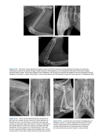
Figure 32.31  The Salter–Harris classification system is used to describe fractures involving the physis. Five types are commonly
            recognized. In general, the greater the type number, the poorer the prognosis as more damage has been done to the physis. Images
            (a) and (b) show a Salter–Harris Type I fracture of the distal femur. The fracture has separated the epiphysis from the metaphysis through
            the physis. In (c) there is a Salter–Harris Type II fracture of the distal femur. The asterisk (*) shows the small segment of metaphyseal bone.


            Figure 32.32  Lateral (a) and ventrodorsal (b) radiographs of
            the pelvis of a mature cat with history of chronic lameness in   Figure 32.33  Lateral (a) and ventrodorsal (b) radiographs of
            the left rear limb. The left femur is shorter than the right and   the pelvis. There is a chronic fracture of the right ilium with
            displays an abnormal shape with chronic cortical remodeling on   medial displacement. The rounded fracture margins with
            the caudal aspect. No soft tissue swelling is present and even   smooth cortical outlines are consistent with chronicity.
            though the cortical margins are irregular, they are intact and   An ischial tuberosity avulsion fragment is also evident.
            sharply marginated. These changes are consistent with a chronic
            healed fracture of the distal femur indicative of previous trauma.