
Page 581 - Feline diagnostic imaging
P. 581
32.2 Accuired one Disease 595
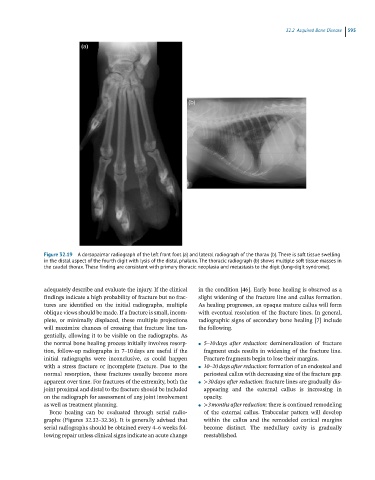
Figure 32.19 A dorsopalmar radiograph of the left front foot (a) and lateral radiograph of the thorax (b). There is soft tissue swelling
in the distal aspect of the fourth digit with lysis of the distal phalanx. The thoracic radiograph (b) shows multiple soft tissue masses in
the caudal thorax. These finding are consistent with primary thoracic neoplasia and metastasis to the digit (lung-digit syndrome).
adequately describe and evaluate the injury. If the clinical in the condition [46]. Early bone healing is observed as a
findings indicate a high probability of fracture but no frac- slight widening of the fracture line and callus formation.
tures are identified on the initial radiographs, multiple As healing progresses, an opaque mature callus will form
oblique views should be made. If a fracture is small, incom- with eventual resolution of the fracture lines. In general,
plete, or minimally displaced, these multiple projections radiographic signs of secondary bone healing [7] include
will maximize chances of crossing that fracture line tan- the following.
gentially, allowing it to be visible on the radiographs. As
the normal bone healing process initially involves resorp- ● 5–10 days after reduction: demineralization of fracture
tion, follow-up radiographs in 7–10 days are useful if the fragment ends results in widening of the fracture line.
initial radiographs were inconclusive, as could happen Fracture fragments begin to lose their margins.
with a stress fracture or incomplete fracture. Due to the ● 10–20 days after reduction: formation of an endosteal and
normal resorption, these fractures usually become more periosteal callus with decreasing size of the fracture gap.
apparent over time. For fractures of the extremity, both the ● >30 days after reduction: fracture lines are gradually dis-
joint proximal and distal to the fracture should be included appearing and the external callus is increasing in
on the radiograph for assessment of any joint involvement opacity.
as well as treatment planning. ● >3 months after reduction: there is continued remodeling
Bone healing can be evaluated through serial radio- of the external callus. Trabecular pattern will develop
graphs (Figures 32.32–32.36). It is generally advised that within the callus and the remodeled cortical margins
serial radiographs should be obtained every 4–6 weeks fol- become distinct. The medullary cavity is gradually
lowing repair unless clinical signs indicate an acute change reestablished.