
Page 576 - Feline diagnostic imaging
P. 576
590 32 Overview of the Musculoskeletal System
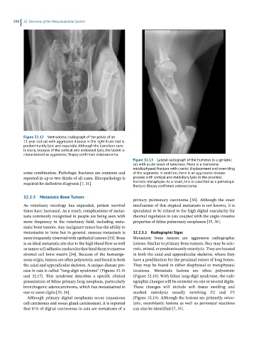
Figure 32.12 Ventrodorsal radiograph of the pelvis of an
11-year-old cat with aggressive disease in the right ilium that is
predominantly lytic and expansile. Although the transition zone
is sharp, because of the cortical and endosteal lysis, the lesion is
characterized as aggressive. Biopsy confirmed osteosarcoma.
Figure 32.13 Lateral radiograph of the humerus in a geriatric
cat with acute onset of lameness. There is a transverse
middiaphyseal fracture with cranial displacement and overriding
some combination. Pathologic fractures are common and of the segments. In addition, there is an aggressive disease
reported in up to two-thirds of all cases. Histopathology is process with cortical and medullary lysis in the proximal
required for definitive diagnosis [7, 31]. humeral metaphysis. As a result, this is classified as a pathologic
fracture. Biopsy confirmed osteosarcoma.
32.2.3 Metastatic Bone Tumors
primary pulmonary carcinoma [36]. Although the exact
As veterinary oncology has expanded, patient survival mechanism of this atypical metastasis is not known, it is
times have increased. As a result, complications of metas- speculated to be related to the high digital vascularity for
tasis commonly recognized in people are being seen with thermal regulation in cats coupled with the angio-invasive
more frequency in the veterinary field, including meta- properties of feline pulmonary neoplasms [35, 36].
static bone tumors. Any malignant tumor has the ability to
metastasize to bone but in general, osseous metastasis is 32.2.3.1 Radiographic Signs
more frequently observed with epithelial tumors [33]. Bone Metastatic bone tumors are aggressive radiographic
is an ideal metastatic site due to the high blood flow as well lesions. Similar to primary bone tumors, they may be scle-
as tumor cell adhesive molecules that bind them to marrow rotic, mixed, or predominantly osteolytic. They are located
stromal cell bone matrix [34]. Because of the hematoge- in both the axial and appendicular skeleton, where they
nous origin, lesions are often polyostotic and found in both have a predilection for the proximal extent of long bones.
the axial and appendicular skeleton. A unique disease pro- They may be found in either diaphyseal or metaphyseal
cess in cats is called “lung-digit syndrome” (Figures 32.16 locations. Metastatic lesions are often polyostotic
and 32.17). This syndrome describes a specific clinical (Figure 32.18). With feline lung-digit syndrome, the radi-
presentation of feline primary lung neoplasia, particularly ographic changes will be centered on one or several digits.
bronchogenic adenocarcinoma, which has metastasized to These changes will include soft tissue swelling and
one or more digits [35, 36]. marked osteolysis usually involving P2 and P3
Although primary digital neoplasms occur (squamous (Figure 32.19). Although the lesions are primarily osteo-
cell carcinoma and sweat gland carcinomas), it is reported lytic, osteoblastic lesions as well as periosteal reactions
that 87% of digital carcinomas in cats are metastases of a can also be identified [7, 35].