
Page 572 - Feline diagnostic imaging
P. 572
586 32 Overview of the Musculoskeletal System
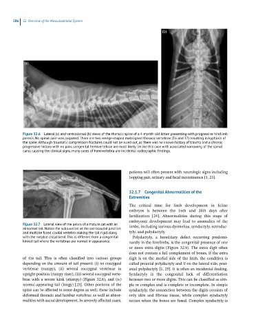
Figure 32.6 Lateral (a) and ventrodorsal (b) views of the thoracic spine of a 4-month-old kitten presenting with progressive hindlimb
paresis. No spinal pain was palpated. There are two wedge-shaped malaligned thoracic vertebrae (T6 and T7) resulting in kyphosis of
the spine. Although traumatic compression fractures could not be ruled out, as there was no known history of trauma and a chronic
progressive history with no pain, congenital hemivertebrae are most likely. Unlike this case with associated narrowing of the spinal
canal causing the clinical signs, many cases of hemivertebra are incidental radiographic findings.
patients will often present with neurologic signs including
hopping gait, urinary and fecal incontinence [1, 23].
32.1.7 Congenital Abnormalities of the
Extremities
The critical time for limb development in feline
embryos is between the 16th and 28th days after
fertilization [24]. Abnormalities during this stage of
embryonic development may lead to anomalies of the
Figure 32.7 Lateral view of the pelvis of a mature cat with an limbs, including various dysmelias, syndactyly, ectrodac-
abnormal tail. Notice the subluxation at the sacrocaudal junction
and multiple fused caudal vertebra making the tail rigid along tyly, and polydactyly.
with the notable distal bend. This is different from a congenital Polydactyly, a hereditary defect occurring predomi-
kinked tail where the vertebrae are normal in appearance. nantly in the forelimbs, is the congenital presence of one
or more extra digits (Figure 32.9). The extra digit often
does not contain a full complement of bones. If the extra
of the tail. This is often classified into various groups digit is on the medial side of the limb, the condition is
depending on the amount of tail present: (i) no coccygeal called preaxial polydactyly and if on the lateral side, post-
vertebrae (rumpy), (ii) several coccygeal vertebrae in axial polydactyly [1, 25]. It is often an incidental finding.
upright position (rumpy riser), (iii) several coccygeal verte- Syndactyly is the congenital lack of differentiation
brae with a severe kink (stumpy) (Figure 32.8), and (iv) between two or more digits. This can be classified as sim-
normal-appearing tail (longy) [23]. Other portions of the ple or complex and is complete or incomplete. In simple
spine can be affected to some degree as well; these include syndactyly, the connection between the digits consists of
deformed thoracic and lumbar vertebrae as well as abnor- only skin and fibrous tissue, while complex syndactyly
malities with sacral development. In severely affected cases, occurs when the bones are fused. Complex syndactyly is