
Page 578 - Feline diagnostic imaging
P. 578
592 32 Overview of the Musculoskeletal System
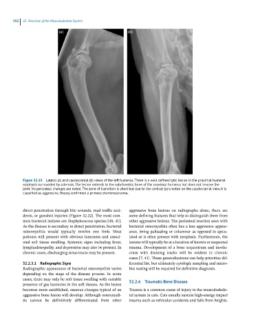
Figure 32.15 Lateral (a) and caudocranial (b) views of the left humerus. There is a well-defined lytic lesion in the proximal humeral
epiphysis surrounded by sclerosis. The lesion extends to the subchondral bone of the proximal humerus but does not involve the
joint. No periosteal changes are noted. The zone of transition is short but due to the cortical lysis noted on the caudocranial view, it is
classified as aggressive. Biopsy confirmed a primary chondrosarcoma.
direct penetration through bite wounds, road traffic acci- aggressive bone lesions on radiographs alone, there are
dents, or gunshot injuries (Figure 32.22). The most com- some defining features that help to distinguish them from
mon bacterial isolates are Staphylococcus species [40, 41]. other aggressive lesions. The periosteal reaction seen with
As the disease is secondary to direct penetration, bacterial bacterial osteomyelitis often has a less aggressive appear-
osteomyelitis would typically involve one limb. Most ance, being palisading or columnar as opposed to spicu-
patients will present with obvious lameness and associ- lated as is often present with neoplasia. Furthermore, the
ated soft tissue swelling. Systemic signs including fever, lesions will typically be at a location of known or suspected
lymphadenopathy, and depression may also be present. In trauma. Development of a bone sequestrum and involu-
chronic cases, discharging sinus tracts may be present. crum with draining tracks will be evident in chronic
cases [7, 41]. These generalizations can help prioritize dif-
32.2.5.1 Radiographic Signs ferential list, but ultimately cytologic sampling and micro-
Radiographic appearance of bacterial osteomyelitis varies bial testing will be required for definitive diagnosis.
depending on the stage of the disease process. In acute
cases, there may only be soft tissue swelling with variable
presence of gas lucencies in the soft tissues. As the lesion 32.2.6 Traumatic Bone Disease
becomes more established, osseous changes typical of an Trauma is a common cause of injury to the musculoskele-
aggressive bone lesion will develop. Although osteomyeli- tal system in cats. Cats usually sustain high-energy impact
tis cannot be definitively differentiated from other trauma such as vehicular accidents and falls from heights.