
Page 583 - Feline diagnostic imaging
P. 583
32.2 Accuired one Disease 597
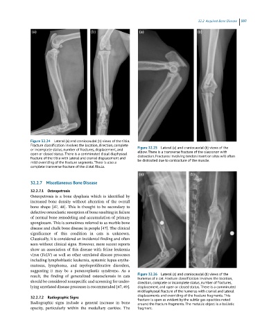
Figure 32.24 Lateral (a) and craniocaudal (b) views of the tibia.
Fracture classification involves the location, direction, complete Figure 32.25 Lateral (a) and craniocaudal (b) views of the
or incomplete status, number of fractures, displacement, and elbow. There is a transverse fracture of the olecranon with
open or closed status. There is a comminuted distal diaphyseal distraction. Fractures involving tendon insertion sites will often
fracture of the tibia with lateral and cranial displacement and be distracted due to contracture of the muscle.
mild overriding of the fracture segments. There is also a
complete transverse fracture of the distal fibula.
32.2.7 Miscellaneous Bone Disease
32.2.7.1 Osteopetrosis
Osteopetrosis is a bone dysplasia which is identified by
increased bone density without alteration of the overall
bone shape [47, 48]. This is thought to be secondary to
defective osteoclastic resorption of bone resulting in failure
of normal bone remodeling and accumulation of primary
spongiosum. This is sometimes referred to as marble bone
disease and chalk bone disease in people [47]. The clinical
significance of this condition in cats is unknown.
Classically, it is considered an incidental finding and often
seen without clinical signs. However, more recent reports
show an association of this disease with feline leukemia
virus (FeLV) as well as other unrelated disease processes
including lymphoblastic leukemia, systemic lupus erythe-
matosus, lymphoma, and myeloproliferative disorders,
suggesting it may be a paraneoplastic syndrome. As a
result, the finding of generalized osteosclerosis in cats Figure 32.26 Lateral (a) and craniocaudal (b) views of the
humerus of a cat. Fracture classification involves the location,
should be considered nonspecific and screening for under- direction, complete or incomplete status, number of fractures,
lying unrelated disease processes is recommended [47, 49]. displacement, and open or closed status. There is a comminuted
middiaphyseal fracture of the humerus with cranial and lateral
32.2.7.2 Radiographic Signs displacements and overriding of the fracture fragments. This
fracture is open as evident by the subtle gas opacities noted
Radiographic signs include a general increase in bone around the fracture fragments. The metallic object is a ballistic
opacity, particularly within the medullary cavities. The fragment.