
Page 590 - Feline diagnostic imaging
P. 590
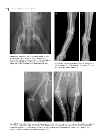
604  32  Overview of the Musculoskeletal System


            Figure 32.39  Classic features of degenerative joint disease
            include sclerosis of the subchondral bone (#), osteophyte
            formation (α), enthesophytes (π), articular mineralization (Ω), and
            joint incongruity(*). All of these features are present in this
            patient with severe osteoarthrosis of the coxofemoral joints.  Figure 32.41  Lateral (a) and dorsoplantar (b) radiographs of
                                                              the tarsus with traumatic luxation of the talocrural joint. This
                                                              was repaired via surgical arthrodesis.


            Figure 32.40  Lateral (a) and craniocaudal (b) radiographs of both stifles in an 11-year-old domestic shorthair that presented with
            acute-onset nonambulatory hindlimb lameness. There is bilateral stifle derangement consistent with complete disruption of the
            ligamentous support structures resulting in luxation. Multiple osseous avulsion fragments are present in both stifles. Surgical
            stabilization was performed with an extracapsular technique.