
Page 593 - Feline diagnostic imaging
P. 593
32.3 oint Disease 607
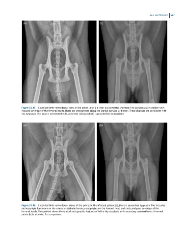
Figure 32.45 Extended limb ventrodorsal view of the pelvis (a) of a 6-year-old domestic shorthair. The acetabula are shallow with
reduced coverage of the femoral heads. There are osteophytes along the cranial acetabular border. These changes are consistent with
hip dysplasia. This case is considered mild. A normal radiograph (b) is provided for comparison.
Figure 32.46 Extended limb ventrodorsal views of the pelvis. In the affected patient (a), there is severe hip dysplasia. This includes
enthesophyte formation on the cranial acetabular border, osteophytes on the femoral head and neck, and poor coverage of the
femoral heads. This patient shows the typical radiographic features of feline hip dysplasia with secondary osteoarthrosis. A normal
pelvis (b) is provided for comparison.