
Page 596 - Feline diagnostic imaging
P. 596
610 32 Overview of the Musculoskeletal System
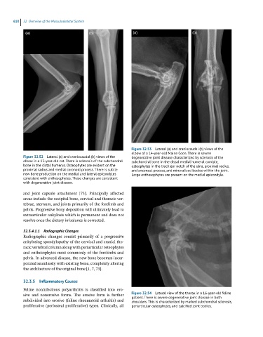
Figure 32.53 Lateral (a) and craniocaudal (b) views of the
elbow of a 14-year-old Maine Coon. There is severe
Figure 32.52 Lateral (a) and craniocaudal (b) views of the degenerative joint disease characterized by sclerosis of the
elbow in a 13-year-old cat. There is sclerosis of the subchondral subchondral bone in the distal medial humeral condyle,
bone in the distal humerus. Osteophytes are evident on the osteophytes in the trochlear notch of the ulna, proximal radius,
proximal radius and medial coronoid process. There is subtle and anconeal process, and mineralized bodies within the joint.
new bone production on the medial and lateral epicondyles Large enthesophytes are present on the medial epicondyle.
consistent with enthesophytes. These changes are consistent
with degenerative joint disease.
and joint capsule attachment [73]. Principally affected
areas include the occipital bone, cervical and thoracic ver-
tebrae, sternum, and joints primarily of the forelimb and
pelvis. Progressive bony deposition will ultimately lead to
extraarticular ankylosis which is permanent and does not
resolve once the dietary imbalance is corrected.
32.3.4.1.1 Radiographic Changes
Radiographic changes consist primarily of a progressive
ankylosing spondylopathy of the cervical and cranial tho-
racic vertebral column along with periarticular osteophytes
and enthesophytes most commonly of the forelimbs and
pelvis. In advanced disease, the new bone becomes incor-
porated seamlessly with existing bone, completely altering
the architecture of the original bone [1, 7, 73].
32.3.5 Inflammatory Causes
Feline noninfectious polyarthritis is classified into ero-
sive and nonerosive forms. The erosive form is further Figure 32.54 Lateral view of the thorax in a 16-year-old feline
patient. There is severe degenerative joint disease in both
subdivided into erosive (feline rheumatoid arthritis) and shoulders. This is characterized by marked subchondral sclerosis,
proliferative (periosteal proliferative) types. Clinically, all periarticular osteophytes, and calcified joint bodies.