
Page 601 - Feline diagnostic imaging
P. 601
32.3 oint Disease 615
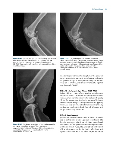
Figure 32.61 Lateral radiograph of the stifle with a small focal Figure 32.63 Large well-developed osseous body in the
area of mineralization likely within the meniscus. This is a cranial aspect of the stifle. This osseous body has characteristics
common finding in cats with an estimated prevalence of of mature bone with cortical and medullary components. This is
36–46%. These are typically confined to the cranial horn of the most consistent with a synovial osteochondroma. This condition
medial meniscus. begins with reactive metaplasia of the synovium with
subsequent formation of an osteochondral nodule in the
synovial lining.
condition begins with reactive metaplasia of the synovium
giving rise to the formation of osteochondral nodules in
the synovial linings. In feline patients, single or multiple
joints can be involved, with the elbow and stifle reported
most frequently [88, 89].
32.3.6.2.1 Radiographic Signs (Figures 32.63–32.65)
Radiographic appearance of a mineralized synovial osteo-
chondroma varies. The lesions are usually well-defined,
rounded, often multiple intraarticular nodules of opacity
[7]. As the lesions often develop in osteoarthritic joints,
concurrent signs of degenerative joint disease are typically
present. As early synovial osteochondromas are primarily
cartilage and poorly mineralized, they will silhouette with
the synovium and synovial fluid.
32.3.6.3 Joint Neoplasia
Synovial cell sarcoma is a rare tumor in cats but is consid-
ered to be the most common primary joint tumor [90].
Synovial neoplasms arise from primitive mesenchymal
Figure 32.62 Focal area of meniscal mineralization noted in precursor cells of the synovial membranes of joints, bursa,
the cranial horn of the meniscus. There is concurrent and tendon sheath [7, 32, 91]. Affected cats will present
degenerative joint disease. The cause of the mineralization is with a soft tissue mass in the vicinity of a joint with
debated, but it appears to be associated with medial
compartment disease in cats. reported cases described in the elbow, carpus, and tarsus