
Page 599 - Feline diagnostic imaging
P. 599
32.3 oint Disease 613
Figure 32.58 Lateral (a) and dorsoplantar (b) views of the
tarsus. There is severe soft tissue swelling of the tarsus in a
juvenile domestic longhair patient secondary to bite wounds. No
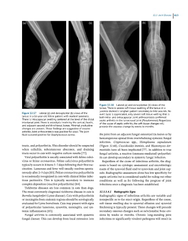
Figure 32.57 Lateral (a) and dorsoplantar (b) views of the overt lysis is appreciated, only severe soft tissue welling that is
tarsus in a 16-year-old feline patient with marked lameness. both intra- and extracapsular. Joint arthrocentesis confirmed
There is intracapsular swelling centered at the level of the distal septic arthritis in the tarsocrural joint (Pseudomonas). Regardless
intertarsal joint. There is osteolysis involving the central, fourth, of the cause of septic arthritis, the soft tissue changes will
and adjacent second and third tarsal bones. Minimal productive precede the osseous change by weeks to months.
changes are present. These findings are suggestive of erosive
arthritis. Joint arthrocentesis was positive for cocci. The joint
fluid cultured positive for Staphylococcus aureus. the joint from an adjacent fungal osteomyelitis lesion or by
hematogenous spread from overwhelming systemic fungal
infection. Cryptococcus spp., Histoplasma capsulatum
tracts, and polyarthritis. This disorder should be suspected (Figure 32.60), Coccidioides immitis, and Blastomyces der-
when cellulitis, subcutaneous abscesses, and draining matitidis have all been implicated [77]. In addition to true
tracts occur in cats with negative culture results [77]. fungal arthritis, a reactive (immune-mediated) polyarthri-
Viral polyarthritis is usually associated with feline calici- tis can develop secondary to systemic fungal infection.
virus or feline coronavirus. Feline calicivirus polyarthritis Regardless of the cause of infectious arthritis, the diag-
typically occurs in kittens 5–7 days following their first vac- nosis is based on cytologic assessment and microbiologic
cination. Lameness and fever will usually resolves sponta- exam of the synovial fluid and/or synovium and joint cap-
neously after 2–5 days [83]. Feline coronavirus polyarthritis sule. Radiographic assessment alone has low specificity for
is occasionally recognized in cats with clinical feline infec- septic arthritis but is considered useful for ruling out other
tious peritonitis. This is typically secondary to immune conditions as well as for following the progress of joint
complex deposition (reactive polyarthritis) [84]. infections once a diagnosis has been established.
Tickborne diseases are less common in cats than dogs.
The most commonly diagnosed tickborne disease in cats is 32.3.5.4.1 Radiographic Signs
Borrelia burgdorferi (Lyme disease). Cats with polyarthritis Radiographic signs of infectious arthritis are variable and
or meningitis from endemic regions should be serologically nonspecific as to the exact origin. Regardless of the cause,
evaluated for Lyme borreliosis. Cats may present with signs soft tissue swelling due to synovial effusion and synovial
of polyarticular lameness, synovitis, meningitis, and sys- thickening is typically present. These changes will precede
temic inflammation [85]. secondary osseous changes such as subchondral bone ero-
Fungal arthritis is commonly associated with systemic sions by weeks or months. Chronic long-standing joint
fungal disease. This can develop from local extension into infections or significantly virulent pathogens will result in