
Page 602 - Feline diagnostic imaging
P. 602
616 32 Overview of the Musculoskeletal System
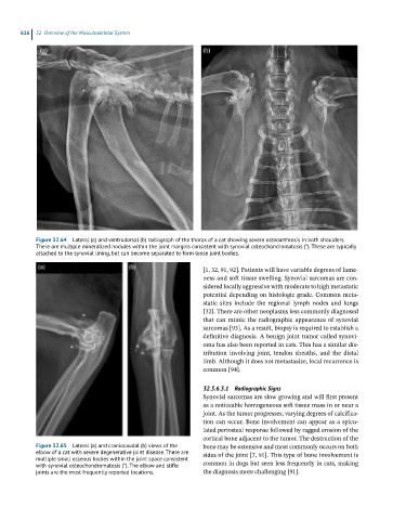
Figure 32.64 Lateral (a) and ventrodorsal (b) radiograph of the thorax of a cat showing severe osteoarthrosis in both shoulders.
There are multiple mineralized nodules within the joint margins consistent with synovial osteochondromatosis (*). These are typically
attached to the synovial lining, but can become separated to form loose joint bodies.
[1, 32, 91, 92]. Patients will have variable degrees of lame -
ness and soft tissue swelling. Synovial sarcomas are con-
sidered locally aggressive with moderate to high metastatic
potential depending on histologic grade. Common meta-
static sites include the regional lymph nodes and lungs
[32]. There are other neoplasms less commonly diagnosed
that can mimic the radiographic appearance of synovial
sarcomas [93]. As a result, biopsy is required to establish a
definitive diagnosis. A benign joint tumor called synovi-
oma has also been reported in cats. This has a similar dis-
tribution involving joint, tendon sheaths, and the distal
limb. Although it does not metastasize, local recurrence is
common [94].
32.3.6.3.1 Radiographic Signs
Synovial sarcomas are slow growing and will first present
as a noticeable homogeneous soft tissue mass in or near a
joint. As the tumor progresses, varying degrees of calcifica-
tion can occur. Bone involvement can appear as a spicu-
lated periosteal response followed by ragged erosion of the
cortical bone adjacent to the tumor. The destruction of the
Figure 32.65 Lateral (a) and craniocaudal (b) views of the bone may be extensive and most commonly occurs on both
elbow of a cat with severe degenerative joint disease. There are sides of the joint [7, 91]. This type of bone involvement is
multiple small osseous bodies within the joint space consistent
with synovial osteochondromatosis (*). The elbow and stifle common in dogs but seen less frequently in cats, making
joints are the most frequently reported locations. the diagnosis more challenging [91].