
Page 598 - Feline diagnostic imaging
P. 598
612 32 Overview of the Musculoskeletal System
antigenic stimulation (reactive polyarthritis) or as a feature
of systemic lupus erythematosus [73, 77]. Radiographic
changes are typically limited to soft tissue swelling and
joint capsule distension. Radiography is primarily used to
distinguish between the erosive and nonerosive forms of
feline polyarthritis. Immune-mediated polyarthritis
(IMPA) is diagnosed in cats with nonerosive polyarthritis
when infectious etiologies are eliminated and there is no
evidence to support systemic lupus erythematosus or reac-
tive polyarthritis. Reactive polyarthritis occurs secondary
to antigenic stimulation from chronic infection, neoplasia,
or drug administration. Infections that have been identi-
fied in cats with presumed reactive polyarthritis include
pneumonia, pyelonephritis, gastrointestinal disease, and
cat bite abscesses.
Systemic lupus erythematosus is an autoimmune disor-
der resulting in immune complex deposition in tissues
leading to inflammation and organ damage. Polyarthritis is
the most commonly reported clinical manifestation of
canine systemic lupus erythematosus but occurs less fre-
quently in cats in which dermatitis, fever, and glomerulo-
nephritis are more frequently reported.
32.3.5.3.1 Radiographic Signs
Radiographic changes are limited to periarticular soft tis-
sue swelling, joint capsule distension, and no overt signs of
subchondral erosions.
32.3.5.4 Infectious Arthritis
Infectious arthritis in cats is much more common than pri-
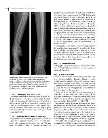
Figure 32.56 Lateral (a) and dorsopalmar (b) views of the
carpus with feline rheumatoid-like polyarthritis (erosive mary immune-mediated disorders. Infectious causes
polyarthritis). Severe subchondral marginal and central osseous include bacteria, bacterial L-forms, Mycoplasma species,
erosions, soft tissue swelling, and periarticular new bone viral causes (calicivirus, feline infectious peritonitis), fungi
production centered at the carpus are visible. Joint tap was (Cryptococcus, Histoplasma), and some tickborne Rickettsia
consistent with suppurative arthritis with negative culture
results typical of this disease process. [77, 79]. Infectious disorders primarily cause arthritis by
direct inoculation in the synovium.
A penetrating bite wound is the most common cause of
32.3.5.2.1 Radiographic Signs (Figure 32.56) septic arthritis. Pasteurella and coliform organisms are
Rheumatoid-like arthritis is characterized by severe sub- most often incriminated. Patients are often systemically ill,
chondral central and marginal erosions along with periar- febrile, and depressed. The affected joint is painful with
ticular soft tissue swelling. Subchondral cyst formation is palpable swelling (Figures 32.57–32.59) [13, 77].
also common. New bone production including osteo- Mycoplasma species are normal inhabitants and gener-
phytes and enthesophytes can occur. In advanced cases, ally considered nonpathogenic. Mycoplasma felis and M.
there is extensive bone destruction resulting in gross gateae have been associated with erosive and nonerosive
deformities of the joint, particularly involving the distal polyarthritis [80–82]. Mycoplasma polyarthritis typically
extremities [7, 77]. results from hematogenous spread of the bacteria to the
joints and is typically described as primarily nonerosive.
32.3.5.3 Nonerosive Immune-Mediated Polyarthritis Screening patients suspected of Mycoplasma polyarthritis
Immune-mediated nonerosive polyarthritis is caused by for underlying immunosuppressive disorders is suggested.
deposition of immune complexes in the synovial mem- Bacterial L-form arthritis is typically secondary to direct
brane with resulting inflammatory response. This can inoculation through bite wounds with the infection spread-
occur secondary to idiopathic disorder, secondary to ing locally and hematogenously to cause cellulitis, draining