
Page 597 - Feline diagnostic imaging
P. 597
32.3 oint Disease 611
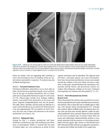
Figure 32.55 Lateral (a) and craniocaudal (b) views of a stifle with degenerative joint disease. There are two small mineralized
opacities in the joint and osteophytes along the distal patella. There is sclerosis evident in the femoral trochlea. The mineralized
opacities may be associated with meniscal mineralization (larger lesion) and an osteochondral fragment (smaller lesion) from cruciate
ligament injury or articular cartilage fragment. Source: Courtesy of Dr Jay Griffin.
forms are similar, with cats appearing stiff, unwilling to capsule attachment may be identified. The adjacent bone
move and resenting any form of handling. Joints are usu- will show a decreased opacity and coarse trabeculation.
ally swollen and painful to palpation. The patient may also Over time, the periosteal proliferation worsens and extends
be febrile and hyporexic. beyond the confines of the joint which can lead to anky-
loses. At this point, marked periarticular osteophytes, sub-
32.3.5.1 Periosteal Proliferative Form chondral cyst-like lesions, and periarticular erosions are
Periosteal proliferative polyarthritis occurs most often in evident with subsequent collapse of the joint. Prominent
male cats but has been reported in female cats as well [74]. enthesopathies are common and often painful [7, 75, 77].
Cats of any age can develop polyarthritis, but it is classi-
cally seen in young adults between the ages of 1 and 5 years 32.3.5.2 Feline Rheumatoid-Like Arthritis
[75–77]. Clinical signs include fever, lethargy, stiffness, (Erosive Form)
swollen, and painful joints particularly of the carpus and Feline rheumatoid-like arthritis is the second form of nonin-
tarsus. Regional lymphadenopathy may also be present. fectious polyarthritis and closely resembles human rheuma-
The stifle, elbow, shoulder, and hip joints are affected to a toid arthritis. This is classically seen in middle-aged to older
lesser extent [78]. Many cats are positive for feline syncytial cats, with Siamese being overrepresented [77]. Although
virus and approximately 50% are positive for FeLV; how- this condition affects multiple joints, the patients often pre-
ever, the exact role of these viral infections in the disease sent with one limb more severely affected. Unlike the peri-
mechanism is not clear [1, 76]. osteal proliferative form, systemic illness and fever typically
do not occur so patients may not present before there are
32.3.5.1.1 Radiographic Signs severe joint deformities. Rheumatoid-like arthritis is com-
During the first 1–2 months, periarticular soft tissue monly associated with the presence of circulating autoanti-
swelling is a predominant sign. The swelling may be either bodies directed against IgG, known as rheumatoid factor
intra- or extracapsular. Within 1–3 months after the onset (RF). Rheumatoid-like arthritis is an aggressive and destruc-
of clinical signs, new bone production at points of joint tive disease that is likely irreversible if not caught early.