
Page 595 - Feline diagnostic imaging
P. 595
32.3 oint Disease 609
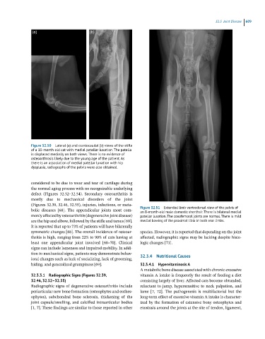
Figure 32.50 Lateral (a) and craniocaudal (b) views of the stifle
of a 10-month-old cat with medial patellar luxation. The patella
is displaced medially on both views. There is no evidence of
osteoarthrosis likely due to the young age of the patient. As
there is an association of medial patellar luxation with hip
dysplasia, radiographs of the pelvis were also obtained.
considered to be due to wear and tear of cartilage during
the normal aging process with no recognizable underlying
defect (Figures 32.52–32.54). Secondary osteoarthritis is
mostly due to mechanical disorders of the joint
(Figures 32.39, 32.46, 32.55), injuries, infections, or meta-
bolic diseases [68]. The appendicular joints most com- Figure 32.51 Extended limb ventrodorsal view of the pelvis of
an 8-month-old male domestic shorthair. There is bilateral medial
monly affected by osteoarthritis (degenerative joint disease) patellar luxation. The coxofemoral joints are normal. There is mild
are the hip and elbow, followed by the stifle and tarsus [69]. medial bowing of the proximal tibia in both rear limbs.
It is reported that up to 73% of patients will have bilaterally
symmetric changes [68]. The overall incidence of osteoar- species. However, it is reported that depending on the joint
thritis is high, ranging from 22% to 90% of cats having at affected, radiographic signs may be lacking despite histo-
least one appendicular joint involved [68–70]. Clinical logic changes [71].
signs can include lameness and impaired mobility. In addi-
tion to mechanical signs, patients may demonstrate behav- 32.3.4 Nutritional Causes
ioral changes such as lack of socializing, lack of grooming,
hiding, and generalized grumpiness [69]. 32.3.4.1 Hypervitaminosis A
A metabolic bone disease associated with chronic excessive
32.3.3.1 Radiographic Signs (Figures 32.39, vitamin A intake is frequently the result of feeding a diet
32.46, 32.52–32.55) consisting largely of liver. Affected cats become obtunded,
Radiographic signs of degenerative osteoarthritis include reluctant to jump, hypersensitive to neck palpation, and
periarticular new bone formation (osteophytes and enthes- lame [7, 72]. The pathogenesis is multifactorial but the
ophytes), subchondral bone sclerosis, thickening of the long-term effect of excessive vitamin A intake is character-
joint capsule/swelling, and calcified intraarticular bodies ized by the formation of extensive bony osteophytes and
[1, 7]. These findings are similar to those reported in other exostosis around the joints at the site of tendon, ligament,