
Page 600 - Feline diagnostic imaging
P. 600
614 32 Overview of the Musculoskeletal System
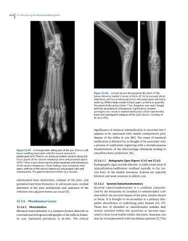
Figure 32.60 Lateral (a) and dorsoplantar (b) views of the
tarsus showing marked erosive arthritis of the tarsocrural, distal
intertarsal, and tarsometatarsal joints with associated soft tissue
swelling. Differentials would include septic arthritis or possibly
rheumatoid-like polyarthritis. Final diagnosis was septic fungal
arthritis secondary to Histoplasma. Significantly virulent
pathogens will result in marked destruction of the subchondral
bone and subsequent collapse of the joint. Source: Courtesy of
Dr Jay Griffin.
significance of meniscal mineralization is uncertain but it
appears to be associated with medial compartment joint
disease of the stifles in cats [86]. The cause of meniscal
ossification is debated but is thought to be associated with
a process of ossification beginning with a chondroosseous
Figure 32.59 A dorsoplantar radiograph of the pes. There is soft transformation of the fibrocartilage ultimately leading to
tissue swelling associated with the second metatarsal cancellous bone production [86].
phalangeal joint. There is an active periosteal reaction along the
distal aspect of the second metatarsal bone and proximal aspect 32.3.6.1.1 Radiographic Signs (Figures 32.61 and 32.62)
of P1. There is lysis involving the distal epiphysis and metaphysis
of the second metatarsus. These findings were consistent with Radiographic signs include discrete, variably sized areas of
septic arthritis of the second metatarsal phalangeal joint and mineralization/ossification confined typically to the cra-
osteomyelitis. The patient had been bitten by a raccoon. nial horn of the medial meniscus. Lesions are typically
bilateral and most common in elderly cats.
subchondral bone destruction, collapse of the joint, and
periosteal new bone formation. In advanced cases, marked 32.3.6.2 Synovial Osteochondromatosis
distortion of the joint architecture and extension of the Synovial osteochondromatosis is a condition character-
infection into adjacent bones can occur [7]. ized by the formation of chondral or osteochondral nod-
ules within the synovial tissues of the joint, tendon sheath
or bursa. It is thought to be secondary to a primary idio-
32.3.6 Miscellaneous Causes
pathic disturbance or underlying joint disease [54, 87].
32.3.6.1 Mineralization These foci of chondral or osteochondral nodules may
Meniscal mineralization is a common feature detected on remain attached within the synovium or become sepa-
conventional orthogonal radiographs of the stifle in domes- rated to form loose bodies within the joints. Burmese cats
tic cats. Estimated prevalence is 36–46%. The clinical may be overrepresented with this disease process [7]. This