
Page 592 - Feline diagnostic imaging
P. 592
606 32 Overview of the Musculoskeletal System
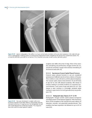
Figure 32.43 Lateral radiographs of the stifles in a 2-year-old domestic shorthair with acute-onset lameness in the right stifle (a).
There is intracapsular swelling present as evidenced by displacement of the infrapatellar fat pad and the caudal facial planes. The
normal left stifle (b) is provided for comparison. Final diagnosis was acute cranial cruciate ligament rupture.
dysplasia also differs from that of dogs. Most of the exten-
sive remodeling and proliferative changes involve the cra-
nial dorsal acetabular margin with minimal remodeling of
the femoral necks [60, 62].
32.3.2.2 Spontaneous Femoral Capital Physeal Fractures
Femoral capital physeal fractures in cats are considered
spontaneous if affected cats do not have a history of trauma.
This condition is also referred to as feline physeal dysplasia.
The normal capital physis closes between 30 and 40 weeks
of age but in cats with physeal dysplasia, this closure is
delayed, leading to increased incidence of atraumatic frac-
tures between 1 and 2 years of age (and older) [63, 64]. This
disease is most common in overweight neutered males
leading to a speculation that early gonadectomy may predis-
pose [65].
32.3.2.2.1 Radiographic Signs (Figures 32.47–32.49)
Radiographic changes include capital physeal incongruity
and displacement of the femoral epiphysis. With chronicity,
Figure 32.44 Two-year-old domestic longhair with acute there will be resorption of the femoral neck and evidence of
lameness localized to the stifle. There is intracapsular swelling, osteolysis, sclerosis, and potentially pseudoarthrosis. This
indicated by the loss of visualization of the infrapatellar fat pad
and caudal displacement of the facial planes. Final diagnosis condition has been reported to be bilateral in up to 63% of
was acute cranial cruciate ligament rupture. cases [63].