
Page 594 - Feline diagnostic imaging
P. 594
608 32 Overview of the Musculoskeletal System
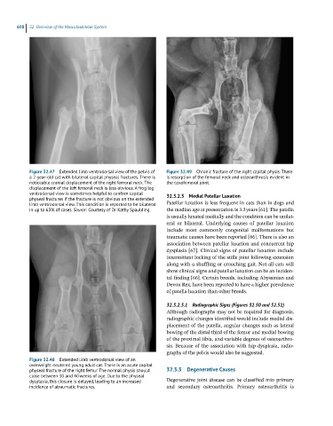
Figure 32.47 Extended limb ventrodorsal view of the pelvis of Figure 32.49 Chronic fracture of the right capital physis. There
a 2-year-old cat with bilateral capital physeal fractures. There is is resorption of the femoral neck and osteoarthrosis evident in
noticeable cranial displacement of the right femoral neck. The the coxofemoral joint.
displacement of the left femoral neck is less obvious. A frog leg
ventrodorsal view is sometimes helpful to confirm capital 32.3.2.3 Medial Patellar Luxation
physeal fractures if the fracture is not obvious on the extended
limb ventrodorsal view. This condition is reported to be bilateral Patellar luxation is less frequent in cats than in dogs and
in up to 63% of cases. Source: Courtesy of Dr Kathy Spaulding. the median age at presentation is 3.3 years [61]. The patella
is usually luxated medially and the condition can be unilat-
eral or bilateral. Underlying causes of patellar luxation
include most commonly congenital malformations but
traumatic causes have been reported [66]. There is also an
association between patellar luxation and concurrent hip
dysplasia [67]. Clinical signs of patellar luxation include
intermittent locking of the stifle joint following extension
along with a shuffling or crouching gait. Not all cats will
show clinical signs and patellar luxation can be an inciden-
tal finding [66]. Certain breeds, including Abyssinian and
Devon Rex, have been reported to have a higher prevalence
of patella luxation than other breeds.
32.3.2.3.1 Radiographic Signs (Figures 32.50 and 32.51)
Although radiographs may not be required for diagnosis,
radiographic changes identified would include medial dis-
placement of the patella, angular changes such as lateral
bowing of the distal third of the femur and medial bowing
of the proximal tibia, and variable degrees of osteoarthro-
sis. Because of the association with hip dysplasia, radio-
graphs of the pelvis would also be suggested.
Figure 32.48 Extended limb ventrodorsal view of an
overweight neutered young adult cat. There is an acute capital
physeal fracture of the right femur. The normal physis should 32.3.3 Degenerative Causes
close between 30 and 40 weeks of age. Due to the physeal
dysplasia, this closure is delayed, leading to an increased Degenerative joint disease can be classified into primary
incidence of atraumatic fractures. and secondary osteoarthritis. Primary osteoarthritis is