
Page 591 - Feline diagnostic imaging
P. 591
32.3 oint Disease 605
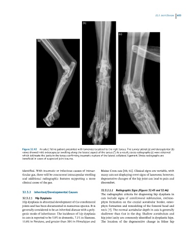
Figure 32.42 An adult feline patient presented with lameness localized to the right tarsus. The survey lateral (a) and dorsoplantar (b)
views showed mild extracapsular swelling along the lateral aspect of the tarsus (*). As a result, stress radiographs (c) were obtained
which delineate the laxity in the tarsus confirming traumatic rupture of the lateral collateral ligament. Stress radiographs are
beneficial in cases of suspected joint trauma.
identified. With traumatic or infectious causes of intraar- Maine Coon cats [60, 61]. Clinical signs are variable, with
ticular gas, there will be concurrent intracapsular swelling many cats not displaying overt signs of lameness; however,
and additional radiographic features supporting a more degenerative changes of the hip joint can lead to pain and
clinical cause of the gas. discomfort.
32.3.2.1.1 Radiographic Signs (Figures 32.45 and 32.46)
32.3.2 Inherited/Developmental Causes
The radiographic criteria for diagnosing hip dysplasia in
32.3.2.1 Hip Dysplasia cats include signs of coxofemoral subluxation, entheso-
Hip dysplasia is abnormal development of the coxofemoral phyte formation on the cranial acetabular border, osteo-
joints and has been documented in numerous species. It is phyte formation and remodeling of the femoral head and
generally considered to be an inherited disease with a poly- neck [7]. The normal acetabular depth in cats is generally
genic mode of inheritance. The incidence of hip dysplasia shallower than that in the dog. Shallow acetabulum and
in cats is reported to be 5.8% in domestic, 7.1% in Siamese, hip joint laxity are commonly identified in dysplastic hips.
15.8% in Persians, and greater than 20% in Himalayan and The location of the degenerative change in feline hip