
Page 2438 - Cote clinical veterinary advisor dogs and cats 4th
P. 2438
Coma 1207
(Continued from previous page)
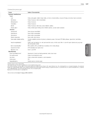
VetBooks.ir Cause † Salient Characteristic
Systemic Noninfectious
Toxins
Lead History, radiographic metallic foreign bodies, red blood cell abnormalities, concurrent GI signs, whole blood lead concentration
Barbiturates History of exposure, blood concentrations
Antidepressants History of exposure
Tranquilizers History of exposure
Alcohol History of exposure, blood levels, severe metabolic acidosis
Ethylene glycol History, ethylene glycol testing, severe metabolic acidosis, calcium oxalate crystalluria
Metabolic
Hypoglycemia* Serum glucose concentration
Hypernatremia Serum sodium concentration
Hyponatremia Serum sodium concentration
Diabetic ketoacidosis Serum and/or urine ketones
Hyperosmolar diabetes mellitus Calculate osmolality (sodium and glucose), predisposing cause of decreased GFR (kidney disease, hypovolemia, heart failure,
shock)
Hepatic encephalopathy* History, serum ammonium, pre- and post-serum bile acids, urine bile acids. Often in cats with hepatic lipidosis and young dogs
with portosystemic shunts.
Uremic encephalopathy* BUN, creatinine, USG, potential history of previous chronic kidney disease
Hypoadrenocorticism ACTH stimulation test, basal cortisol
Myxedema coma Serum T 4 and TSH Differentials, Lists, and Mnemonics
Miscellaneous
Severe hypovolemia/shock* Assessment of physical perfusion parameters, lactate, base excess
Post-cardiac arrest History of cardiorespiratory arrest
Heat stroke History, environmental temperature, rectal temperature
Erythrocytosis PCV
Hyperglobulinemia Globulin levels on chemistry panel
*Common causes of coma in small animals.
† Can evaluate with over-the-counter drug testing kit or human hospital toxicology screen.
ACTH, Adrenocorticotropic hormone; BMBT, buccal mucosal bleeding time; BUN, blood urea nitrogen; CDV, canine distemper virus; CSF, cerebrospinal fluid; CT, computed tomography; GFR, glomerular
filtration rate; GI, gastrointestinal; IMHA, immune-mediated hemolytic anemia; MRI, magnetic resonance imaging; PCV, packed cell volume; PT, prothrombin time; PTT, partial thromboplastin time; T 4 , thyroxine;
TEG, thromboelastography; TSH, thyroid-stimulating hormone; USG, urine specific gravity.
Reproduced from the third edition in unabridged form.
THIRD EDITION AUTHOR: Søren R. Boysen, DVM, DACVECC
www.ExpertConsult.com