Page 2462 - Cote clinical veterinary advisor dogs and cats 4th
P. 2462
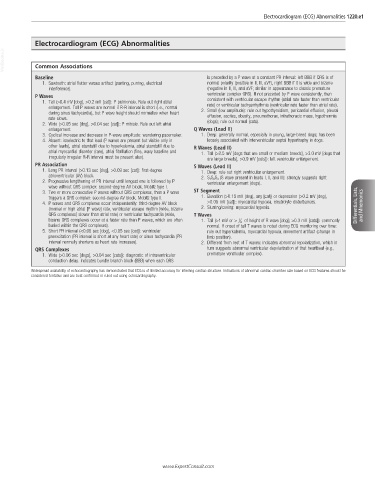
Electrocardiogram (ECG) Abnormalities 1220.e1


            Electrocardiogram (ECG) Abnormalities
  VetBooks.ir  Common Associations


             Baseline                                                is preceded by a P wave at a constant PR interval: left BBB if QRS is of
               1.  Sawtooth: atrial flutter versus artifact (panting, purring, electrical   normal polarity (positive in II, III, aVF), right BBB if it is wide and bizarre
                 interference).                                      (negative in II, III, and aVF; similar in appearance to classic premature
             P Waves                                                 ventricular complex QRS). If not preceded by P wave consistently, then
               1.  Tall (>0.4 mV [dog], >0.2 mV [cat]): P pulmonale. Rule out right atrial   consistent with ventricular escape rhythm (atrial rate faster than ventricular
                 enlargement. Tall P waves are normal if R-R interval is short (i.e., normal   rate) or ventricular tachyarrhythmia (ventricular rate faster than atrial rate).
                 during sinus tachycardia), but P wave height should normalize when heart   2.  Small (low amplitude): rule out hypothyroidism, pericardial effusion, pleural
                 rate slows.                                         effusion, ascites, obesity, pneumothorax, intrathoracic mass, hypothermia
               2.  Wide (>0.05 sec [dog], >0.04 sec [cat]): P mitrale. Rule out left atrial   (dogs); rule out normal (cats).
                 enlargement.                                   Q Waves (Lead II)
               3.  Cyclical increase and decrease in P-wave amplitude: wandering pacemaker.  1.  Deep: generally normal, especially in young, large-breed dogs; has been
               4.  Absent: isoelectric to that lead (P waves are present but visible only in   loosely associated with interventricular septal hypertrophy in dogs.
                 other leads), atrial standstill due to hyperkalemia, atrial standstill due to   R Waves (Lead II)
                 atrial myocardial disorder (rare), atrial fibrillation (fine, wavy baseline and   1.  Tall (>2.5 mV [dogs that are small or medium breeds], >3.0 mV [dogs that
                 irregularly irregular R-R interval must be present also).  are large breeds], >0.9 mV [cats]): left ventricular enlargement.
             PR Association                                     S Waves (Lead II)
               1.  Long PR interval (>0.13 sec [dog], >0.09 sec [cat]): first-degree   1.  Deep: rule out right ventricular enlargement.
                 atrioventricular (AV) block.                      2.  S 1 S 2S 3 (S wave present in leads I, II, and III): strongly suggests right
               2.  Progressive lengthening of PR interval until longest one is followed by P   ventricular enlargement (dogs).
                 wave without QRS complex: second-degree AV block, Mobitz type I.
               3.  Two or more consecutive P waves without QRS complexes, then a P wave   ST Segment
                 triggers a QRS complex: second-degree AV block, Mobitz type II.  1.  Elevation (>0.15 mV [dog], any [cat]) or depression (>0.2 mV [dog],
               4.  P waves and QRS complexes occur independently: third-degree AV block   >0.05 mV [cat]): myocardial hypoxia, electrolyte disturbances.  Differentials, Lists,   and Mnemonics
                 (normal or high atrial [P wave] rate, ventricular escape rhythm [wide, bizarre   2.  Slurring/coving: myocardial hypoxia.
                 QRS complexes] slower than atrial rate) or ventricular tachycardia (wide,   T Waves
                 bizarre QRS complexes occur at a faster rate than P waves, which are often   1.  Tall (>1 mV or >  1 4  of height of R wave [dog]; >0.3 mV [cats]): commonly
                 buried within the QRS complexes).                   normal. If onset of tall T waves is noted during ECG monitoring over time:
               5.  Short PR interval (<0.06 sec [dog], <0.05 sec [cat]): ventricular   rule out hyperkalemia, myocardial hypoxia, movement artifact (change in
                 preexcitation (PR interval is short at any heart rate) or sinus tachycardia (PR   limb position).
                 interval normally shortens as heart rate increases).  2.  Different from rest of T waves: indicates abnormal repolarization, which in
             QRS Complexes                                           turn suggests abnormal ventricular depolarization of that heartbeat (e.g.,
               1.  Wide (>0.06 sec [dogs], >0.04 sec [cats]): diagnostic of intraventricular   premature ventricular complex).
                 conduction delay. Indicates bundle branch block (BBB) when each QRS

           Widespread availability of echocardiography has demonstrated that ECG is of limited accuracy for inferring cardiac structure. Indications of abnormal cardiac chamber size based on ECG features should be
           considered tentative and are best confirmed or ruled out using echocardiography.


                                                      www.ExpertConsult.com

