
Page 2458 - Cote clinical veterinary advisor dogs and cats 4th
P. 2458
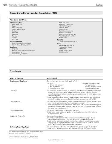
1218  Disseminated Intravascular Coagulation (DIC)                                                   Dysphagia


            Disseminated Intravascular Coagulation (DIC)
  VetBooks.ir  Associated Conditions


            Inflammatory/Toxic                                    Heart base tumor
              Acute hepatic injury                                Intra-abdominal sarcoma
              Sepsis                                              Malignant histiocytosis
              Acute kidney injury                                 Mast cell tumor
              Fever of unknown origin                             Oral carcinoma
              Meningitis                                          Perianal adenocarcinoma
              Pancreatitis                                        Pulmonary carcinoma
              Pneumonia                                           Nasal squamous cell carcinoma
              Pyometra                                            Squamous papilloma (vulvar)
              Snakebite                                           Bladder tumor
              Heatstroke                                        Parasitic
            Immune-Mediated                                       Angiostrongylus vasorum infection
              Immune-mediated thrombocytopenia                  Other
              Immune-mediated hemolytic anemia                    Hemorrhagic gastroenteritis
            Neoplastic                                            Cold agglutinin disease
              Lymphoma                                            Multitrauma
              Mammary adenocarcinoma/mammary tumor                Gastric dilation/volvulus
              Hemangiosarcoma                                     Diabetes mellitus
              Splenic mass (type unknown)


            Dysphagia


            Anatomic Location                       Key Feature(s)
            Oropharyngeal Dysphagia                 Oral examination and observation of attempts to eat/drink
                                                    Oral lesion:                           Pharyngeal/cricopharyngeal lesion:
                                                      •  Difficulty with prehension         •  Normal prehension
                                                      •  Pain on prehension                 •  Repeated attempts to swallow
                                                      •  Food dropping from mouth           •  Choking/gagging common
              Oral stage                            Ddx: oral masses, retrobulbar abscess, FB, trauma (e.g., mandibular/maxillary fracture), TMJ disorders
                                                      (luxation, fracture, craniomandibular osteopathy), dental pain, glossitis, stomatitis, cleft palate,
                                                      lingual frenulum lesion, masticatory myositis, myopathy, cranial nerve V, VII, and XII dysfunction (any
                                                      cause, including rabies)
                                                    Consider: sedated/anesthetized oral exam, dental radiographs, survey skull/neck radiographs (special
                                                      attention to mandibles, temporomandibular joint and teeth), advanced imaging (CT or MRI)
              Pharyngeal stage                      Ddx: pharyngeal inflammation (trauma, abscess, eosinophilic granuloma, pharyngeal sialocele), cranial
                                                      nerve V, VII, IX, and X dysfunction, FB, lymphadenopathy, neoplasia
                                                    Consider: palpate pharynx and neck for masses, asymmetry, or pain; sedated/anesthetized pharyngeal
                                                      examination; pharyngeal region radiographs to rule out mass lesions, FB; barium contrast and video
                                                      fluoroscopy (pharyngeal dysfunction)
              Cricopharyngeal stage                 Ddx: cricopharyngeal achalasia, cricopharyngeal asynchrony
                                                    Consider: breed (cocker and springer spaniels, toy breeds, golden retrievers), differentiation via
                                                      fluoroscopic contrast study
            Esophageal Dysphagia                    Characterized by regurgitation
                                                    Ddx: primary (idiopathic) megaesophagus, secondary megaesophagus, esophageal stricture,
                                                      extraesophageal compression (e.g., vascular ring anomaly), esophageal diverticulum
                                                    Consider: cervical and thoracic radiograph (megaesophagus and to rule out aspiration pneumonia),
                                                      esophagoscopy for esophagitis, FB (which can be retrieved) and strictures (which can be balloon-
                                                      dilated), specific tests for cause of megaesophagus (if present)
            Gastroesophageal Dysphagia              Ddx: reflux esophagitis, hiatal hernia, periesophageal hernia, gastroesophageal intussusception
                                                    Consider: thoracic and abdominal radiographs, abdominal ultrasound, flouroscopy, endoscopy
           Ddx, Differential diagnosis; FB, foreign body; TMJ, temporomandibular joint.
           Reproduced from the third edition in modified form.
           THIRD EDITION AUTHOR: Oriana D. Raab, DVM, DACVIM
                                                     www.ExpertConsult.com