
Page 2541 - Cote clinical veterinary advisor dogs and cats 4th
P. 2541
Regurgitation 1277
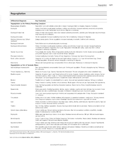
Regurgitation
VetBooks.ir Differential Diagnosis Key Feature(s)
Regurgitation as the Primary Presenting Complaint
Megaesophagus (idiopathic) Regurgitation can be immediately postprandial or delayed. Esophageal dilation on imaging. Diagnosis of exclusion.
Esophagitis History of risk factors (recent general anesthesia; vomiting; oral antibiotics). Increased swallowing movements and salivation.
Inappetence due to pain. Radiographs may be normal. Diagnosis on endoscopy.
Esophageal foreign body History of foreign body ingestion. Acute onset increased swallowing movements, salivation, pain. Radiographs may show foreign
body. Endoscopy for confirmation.
Esophageal neoplasia Chronic regurgitation. Increased swallowing movements. Pain on swallowing. Endoscopy for diagnosis.
Infectious (Spirocerca lupi; Pythium Enlarged salivary glands. Chronic regurgitation. Increased swallowing movements. Confirmed with endoscopy.
insidiosum)
Esophageal diverticulum Partial dilation seen on contrast radiographs or endoscopy.
Esophageal stricture/stenosis History of risk factors (recent general anesthesia; vomiting; oral antibiotics; foreign body removed). Increased swallowing
movements and salivation. Inappetence due to pain. Regurgitation immediate after eating. Liquids kept down. Contrast
radiographs or endoscopy is diagnostic.
Vascular ring anomaly Young animals after weaning. History of regurgitation starting shortly after introduction of solids. Usually ravenous appetite but
poor growth. Diagnoses made on plain dorsoventral thoracic radiograph.
Pharyngeal obstruction Increased swallowing movements. Pawing at throat. Inappetence. Endoscopy may help but fluoroscopy best.
Pyloric outflow obstruction Congenital: 4-12 months old. Boston terriers and English bulldogs. Acquired: 4- to 7-year-old small-breed dogs. Chronic
vomiting/regurgitation. Ultrasound; contrast radiographs or endoscopy for diagnosis.
Hiatal hernia Shar-pei and brachycephalic dogs overrepresented. Chronic clinical signs. Fluoroscopy or endoscopy for diagnosis. Differentials, Lists, and Mnemonics
Regurgitation as Part of Systemic Illness
Gastroesophageal intussusception Rare. German shepherds overrepresented. Severe pain. Vomiting and regurgitation. Thoracic radiographs may help. Endoscopy
for confirmation.
Thoracic neoplasia (e.g., thymoma) Usually over 8 years of age. Dyspnea. Hypercalcemia often present. Thoracic radiographs show cranial mediastinal mass.
Myasthenia gravis Congenital <8 weeks of age in Jack Russell and smooth fox terriers. Acquired—German shepherds, golden retrievers, German
short-haired pointers, others. In cats, +/− history of methimazole treatment. Generalized limb weakness (worse with exercise)
often present. Can cause regurgitation alone. Acetylcholine receptor antibody test for confirmation.
Botulism Possible history of ingestion of contaminated food or carrion. Acute and rapid generalized weakness. Tail wag is maintained.
Tetanus History of wound or teething. Sensitive to touch and noise. Muscle spasm of the face and jaw. Rigid paresis. Mild hyperthermia.
Organophosphate intoxication Acute. Vomiting/regurgitation and diarrhea. Generalized weakness. Muscle tremors. Miosis. Acetylcholinesterase levels for
confirmation.
Dysautonomia Mostly young adults. Vomiting/regurgitation; diarrhea, dysuria, mydriasis, coughing and nasal discharge may be present. Ocular
pilocarpine test rules out anticholinergic intoxication. Confirmation: histopathologic exam of autonomic ganglia.
Polyradiculoneuritis Lower motor neuron paralysis beginning in hindlimbs and progressing to full paralysis within 10 days. Nociception maintained
+/− enhanced.
Tick paralysis Finding a tick or tick crater. Hindlimb weakness with progression to generalized weakness—usually acute. Tail wag preserved.
Voice change and nystagmus may be present. Dysphagia. Rapid (hours) improvement in signs with tick removal.
Lead History of potential or confirmed exposure. Neurologic signs, vomiting, diarrhea, abdominal pain. Hypochromic anemia. Signs can
be acute or chronic. Blood lead level for confirmation.
Thallium Rare. Vomiting; diarrhea; regurgitation. Alopecia. Neurologic deficits include tremors and ataxia. Thallium concentrations in hair
and blood can confirm.
Systemic lupus erythematosus Young to middle age. Skin and oral mucosal lesions. Numerous other immune-mediated conditions: polyarthritis; hemolytic
anemia; thrombocytopenia. A positive ANA titer.
Polymyositis Middle-aged large-breed dogs. Acute or chronic signs. Weakness worsens with exercise. Stiff gait. EMG and muscle biopsies
can help confirm.
Dermatomyositis Young <1-year-old dogs. Collies and Shetland sheepdogs. Skin lesions on extremities. Muscle atrophy. Weakness and lameness.
Painful and inappetent. Skin and muscle biopsies diagnostic.
Hypoadrenocorticism Young to middle-aged dogs. Weakness and lethargy with anorexia and vomiting often wax and wane. Suspect if hyperkalemia
and hyponatremia. Lack of stress leukogram (eosinophilia). ACTH stimulation test for confirmation.
Hypothyroidism Other presenting signs include mental dullness, weakness and exercise intolerance. Seborrhea and bilateral symmetrical hair
loss with a “rat tail.” Measure T 4 in conjunction with TSH.
ACTH, Adrenocorticotropic hormone; ANA, antinuclear antibody; EMG, electromyogram; T 4 , thyroxine; TSH, thyroid-stimulating hormone.
Reproduced from the third edition in modified form.
THIRD EDITION AUTHOR: Ninette Keller, BVSc, MMedVet
www.ExpertConsult.com