
Page 2545 - Cote clinical veterinary advisor dogs and cats 4th
P. 2545
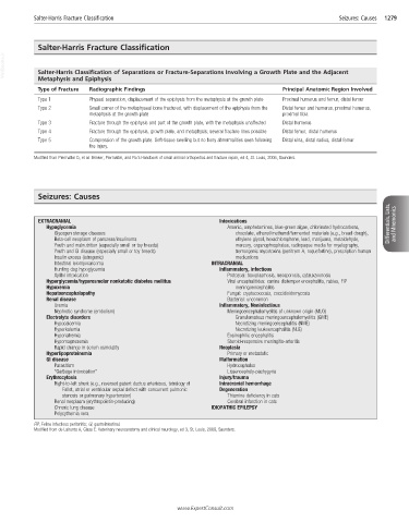
Salter-Harris Fracture Classification                                                       Seizures: Causes   1279


            Salter-Harris Fracture Classification
  VetBooks.ir  Salter-Harris Classification of Separations or Fracture-Separations Involving a Growth Plate and the Adjacent


            Metaphysis and Epiphysis
            Type of Fracture  Radiographic Findings                                   Principal Anatomic Region Involved
            Type 1          Physeal separation, displacement of the epiphysis from the metaphysis at the growth plate  Proximal humerus and femur, distal femur
            Type 2          Small corner of the metaphyseal bone fractured, with displacement of the epiphysis from the   Distal femur and humerus, proximal humerus,
                            metaphysis at the growth plate                            proximal tibia
            Type 3          Fracture through the epiphysis and part of the growth plate, with the metaphysis unaffected  Distal humerus
            Type 4          Fracture through the epiphysis, growth plate, and metaphysis; several fracture lines possible  Distal femur, distal humerus
            Type 5          Compression of the growth plate. Soft-tissue swelling but no bony abnormalities seen following   Distal ulna, distal radius, distal femur
                            the injury.
           Modified from Piermattei D, et al: Brinker, Piermattei, and Flo’s Handbook of small animal orthopedics and fracture repair, ed 4, St. Louis, 2006, Saunders.


            Seizures: Causes


             EXTRACRANIAL                                          Intoxications                                      Differentials, Lists,
               Hypoglycemia                                          Arsenic, amphetamines, blue-green algae, chlorinated hydrocarbons,   and Mnemonics
                 Glycogen storage diseases                             chocolate, ethanol/methanol/fermented materials (e.g., bread dough),
                 Beta-cell neoplasm of pancreas/insulinoma             ethylene glycol, hexachlorophene, lead, marijuana, metaldehyde,
                 Youth and malnutrition (especially small or toy breeds)  mercury, organophosphates, radiopaque media for myelography,
                 Youth and GI disease (especially small or toy breeds)  tremorgenic mycotoxins (penitrem A, roquefortine), prescription human
                 Insulin excess (iatrogenic)                           medications
                 Intestinal leiomyosarcoma                      INTRACRANIAL
                 Hunting dog hypoglycemia                          Inflammatory, Infectious
                 Xylitol intoxication                                Protozoal: toxoplasmosis, neosporosis, cytauxzoonosis
               Hyperglycemia/hyperosmolar nonketotic diabetes mellitus  Viral encephalitides: canine distemper encephalitis, rabies, FIP
               Hypoxemia                                               meningoencephalitis
               Hepatoencephalopathy                                  Fungal: cryptococcosis, coccidioidomycosis
               Renal disease                                         Bacterial: uncommon
                 Uremia                                            Inflammatory, Noninfectious
                 Nephrotic syndrome (embolism)                       Meningoencephalomyelitis of unknown origin (MUO)
               Electrolyte disorders                                   Granulomatous meningoencephalomyelitis (GME)
                 Hypocalcemia                                          Necrotizing meningoencephalitis (NME)
                 Hyperkalemia                                          Necrotizing leukoencephalitis (NLE)
                 Hyponatremia                                        Eosinophilic encephalitis
                 Hypomagnesemia                                      Steroid-responsive meningitis-arteritis
                 Rapid change in serum osmolality                  Neoplasia
               Hyperlipoproteinemia                                  Primary or metastatic
               GI disease                                          Malformation
                 Parasitism                                          Hydrocephalus
                 “Garbage intoxication”                              Lissencephaly-pachygyria
               Erythrocytosis                                      Injury/trauma
                 Right-to-left shunt (e.g., reversed patent ductus arteriosus, tetralogy of   Intracranial hemorrhage
                    Fallot, atrial or ventricular septal defect with concurrent pulmonic   Degeneration
                    stenosis or pulmonary hypertension)              Thiamine deficiency in cats
                 Renal neoplasm (erythropoietin-producing)           Cerebral infarction in cats
                 Chronic lung disease                           IDIOPATHIC EPILEPSY
                 Polycythemia vera
           FIP, Feline infectious peritonitis; GI, gastrointestinal.
           Modified from de Lahunta A, Glass E: Veterinary neuroanatomy and clinical neurology, ed 3, St. Louis, 2009, Saunders.


                                                      www.ExpertConsult.com