
Page 1067 - Small Animal Internal Medicine, 6th Edition
P. 1067
CHAPTER 58 Lesion Localization and the Neurologic Examination 1039
Sensory signals
UMN control to brain Dorsal root
VetBooks.ir (sensory)
from brain
Spinal
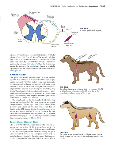
nerve FIG 58.2
A single spinal cord segment.
White Peripheral
matter nerve
Gray
matter
Neuromuscular
Ventral root junction
(motor)
intumescence
intumescence
directed towards the side opposite the lesion (see Vestibular
disease, Chapter 63). Severe lesions of the rostral cerebellum Cervical Lumbar
may result in opisthotonus with rigid extension of all four { {
limbs with flexed hips (decerebellate posture) (see the dis- C1-C5 C6-T2 L4-S2
cussion of posture, p. 1043). Box 58.3 lists the clinical signs T3-L3
caused by lesions of the cerebellum. Causes of cerebellar
dysfunction are discussed with other intracranial disorders
in Chapter 60.
SPINAL CORD
The spinal cord resides entirely within the bony vertebral
column. It is composed of a central H-shaped core of gray
matter surrounded by white matter. Spinal cord gray matter
contains the cell bodies of interneurons and lower motor
neurons (LMNs). White matter is composed of nerve fibers FIG 58.3
organized into columns of ascending and descending long Spinal cord segments at the cervical intumescence (C6-T2)
tracts. These long tracts transmit ascending sensory infor- and the lumbar intumescence (L4-S3) give rise to the
mation (proprioception, touch, temperature, pressure, and important peripheral nerves of the limbs.
pain) and descending motor signals between higher centers
in the brain and spinal cord neurons.
The spinal cord can be functionally divided into seg-
ments, with each spinal cord segment giving rise to one pair
of spinal nerves (left and right), each of which has a dorsal
(sensory) and ventral (motor) root (Fig. 58.2). The cell UMN
bodies for the LMNs supplying the thoracic limbs are in the
ventral gray matter within a thickened region of the cord LMN
called the cervical intumescence (segments C6-T2), whereas
the LMNs supplying the pelvic limbs originate in the lumbar
intumescence (segments L4-S3; Fig. 58.3).
Lower Motor Neuron Signs
The LMN is the efferent neuron that directly connects the
central nervous system (CNS) to a muscle or gland (Fig.
58.4). Components of LMNs include the nerve cell bodies
within the ventral gray matter, the axons leaving the spinal FIG 58.4
canal as ventral nerve roots and spinal nerves, and the The upper motor neuron (UMN) and lower motor neuron
peripheral nerves formed by the spinal nerves that terminate (LMN) systems are responsible for mediating normal motor
at the neuromuscular junction (NMJ) in the muscle to function.