
Page 1069 - Small Animal Internal Medicine, 6th Edition
P. 1069
CHAPTER 58 Lesion Localization and the Neurologic Examination 1041
cord and brainstem to the brain. Most of these tracts ascend BOX 58.4
the ipsilateral spinal cord and cross over in the rostral brain-
VetBooks.ir stem to reach the contralateral cerebrum (see Fig. 58.5). Localization of Spinal Cord Disease
Patients with a unilateral forebrain lesion will typically expe-
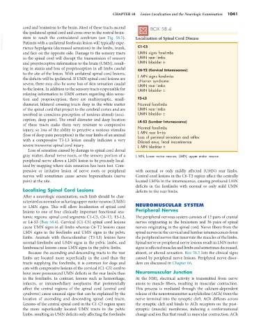
C1-C5
rience hypalgesia (decreased sensation) in the limbs, trunk,
and face on the opposite side. Damage to the sensory tracts UMN signs forelimbs
in the spinal cord will disrupt the transmission of sensory UMN rear limbs
and proprioceptive information to the brain (UMN), result- UMN bladder ±
ing in ataxia and loss of proprioception in all limbs caudal C6-T2 (Cervical Intumescence)
to the site of the lesion. With unilateral spinal cord lesions, L MN signs forelimbs
the deficits will be ipsilateral. If UMN spinal cord lesions are ±Horner syndrome
severe, there may also be some loss of skin sensation caudal UMN rear limbs
to the lesion. In addition to the sensory tracts responsible for UMN bladder ±
relaying information to UMN centers regarding skin sensa-
tion and proprioception, there are multisynaptic, small- T3-L3
diameter, bilateral crossing tracts deep in the white matter Normal forelimbs
of the spinal cord that project to the cerebral cortex and are UMN rear limbs
involved in conscious perception of noxious stimuli (noci- UMN bladder ±
ception, deep pain). The small diameter and deep location L4-S3 (Lumbar Intumescence)
of these tracts make them very resistant to compressive
injury, so loss of the ability to perceive a noxious stimulus Normal forelimbs
L MN rear limbs
(loss of deep pain perception) in the rear limbs of an animal Loss of perineal sensation and reflex
with a compressive T3-L3 lesion usually indicates a very Dilated anus, fecal incontinence
severe transverse spinal cord injury. L MN bladder ±
Loss of sensation caused by damage to spinal cord dorsal
gray matter, dorsal nerve roots, or the sensory portion of a L MN, Lower motor neuron; UMN, upper motor neuron.
peripheral nerve allows a LMN lesion to be precisely local-
ized by mapping where skin sensation has been lost. Com-
pressive or irritative lesion of nerve roots or peripheral with normal or only mildly affected (UMN) rear limbs.
nerves will sometimes cause severe hyperesthesia (nerve Central cord lesions in the C6-T2 region affect the centrally
pain) at the site. located LMNs in the intumescence, causing profound LMN
deficits in the forelimbs with normal or only mild UMN
Localizing Spinal Cord Lesions deficits in the rear limbs.
After a neurologic examination, each limb should be char-
acterized as normal or as having upper motor neuron (UMN)
or LMN signs. This will allow localization of spinal cord NEUROMUSCULAR SYSTEM
lesions to one of four clinically important functional ana- Peripheral Nerves
tomic regions: spinal cord segments C1-C5, C6-T2, T3-L3, The peripheral nervous system consists of 12 pairs of cranial
or L4-S3 (Box 58.4). Cervical (C1-C5) spinal cord lesions nerves originating in the brainstem and 36 pairs of spinal
cause UMN signs in all limbs whereas C6-T2 lesions cause nerves originating in the spinal cord. Nerve fibers from the
LMN signs in the forelimbs and UMN signs in the pelvic spinal nerves in the cervical and lumbar intumescences form
limbs. Animals with thoracolumbar (T3-L3) lesions have the peripheral nerves that innervate the muscles of the limbs.
normal forelimbs and UMN signs in the pelvic limbs, and Spinal nerve or peripheral nerve lesions result in LMN motor
lumbosacral lesions cause LMN signs in the pelvic limbs. signs in affected muscles and limbs and sometimes decreased,
Because the ascending and descending tracts to the rear absent, or altered sensation. Box 58.5 lists the clinical signs
limbs are located more superficially in the cord than the caused by peripheral nerve lesions. Peripheral nerve disor-
tracts supplying the forelimbs, it is common for dogs and ders are discussed in Chapter 66.
cats with compressive lesions of the cervical (C1-C5) cord to
have more pronounced UMN deficits in the rear limbs than Neuromuscular Junction
in the forelimbs. In contrast, lesions such as hemorrhage, At the NMJ, electrical activity is transmitted from nerve
infarcts, or intramedullary neoplasms that preferentially axons to muscle fibers, resulting in muscular contraction.
affect the central regions of the spinal cord (central cord This process is mediated through the calcium-dependent
syndrome) cause unusual signs that can be explained by the release of the neurotransmitter acetylcholine (ACh) from the
location of ascending and descending spinal cord tracts. nerve terminal into the synaptic cleft. ACh diffuses across
Lesions of the central spinal cord in the C1-C5 region spare the synaptic cleft and binds to ACh receptors on the post-
the more superficially located UMN tracts to the pelvic synaptic (muscle) membrane, inducing a conformational
limbs, resulting in UMN deficits only affecting the forelimbs change and ion flux that result in muscular contraction. ACh