
Page 1073 - Small Animal Internal Medicine, 6th Edition
P. 1073
CHAPTER 58 Lesion Localization and the Neurologic Examination 1045
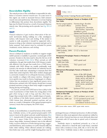
Decerebellate Rigidity TABLE 58.3
The rostral portion of the cerebellum is responsible for inhi-
VetBooks.ir bition of excessive extensor muscle tone. An acute lesion in Localizing Lesions Causing Paresis and Paralysis
this region can result in increased thoracic limb extensor
Tetraparesis/Tetraplegia: Paresis or Paralysis of All
muscle tone and opisthotonus. Mentation is normal, helping
to differentiate this posture from decerebrate rigidity. The Four Limbs
hips may be flexed forward as a result of increased iliopsoas Normal proprioception Nonneurologic disorders
muscle tone. This posturing can be episodic (see Fig. 58.9, B and spinal reflexes (cardiac disease,
and C). hypoglycemia, electrolyte
abnormalities, hypoxemia)
GAIT Myasthenia gravis
Generalized muscle disorders
Clinical evaluation of gait involves observation of the ani-
mal’s movements during walking on a flat, nonslippery LMN fore and rear Generalized disorders of spinal
cord ventral gray matter,
surface, with frequent turns and circling. If the animal is ventral nerve roots, peripheral
unable to walk unassisted, it should be supported with a nerves or neuromuscular
harness or sling so that voluntary movement and gait can be junction
better assessed. Each patient must be evaluated for paresis LMN forelimbs, UMN C6-T2 spinal cord
(weakness), ataxia, lameness, and circling. rear limbs
Paresis/Paralysis UMN forelimbs, UMN C1-C5 or brainstem
rear limbs
Paresis is defined as weakness or inability to support weight
(LMN paresis) or inability to generate a normal gait (UMN Paraparesis/Paraplegia: Paresis or Paralysis of
paresis). Paralysis is the term used to describe the loss of all Rear Limbs
voluntary movement (Table 58.3). When animals are still Normal forelimbs, L4-S3 spinal cord
ambulatory, the gait that results from LMN disease is mark- LMN rear limbs
edly different from the gait that results from a UMN lesion. Normal forelimbs, T3-L3 spinal cord
Animals with LMN disease are usually profoundly weak UMN rear limbs
(paretic), the muscles in affected limbs are relatively flaccid,
and they take small steps, attempting to maintain their feet Monoparesis/Monoplegia: Paresis or Paralysis of
under their center of gravity. Their short-strided, choppy gait One Limb
is commonly mistaken for an orthopedic lameness, and they LMN Lesion of the LMN directly
may tremble or collapse with minor exertion. Attempts to innervating the affected limb
move quickly may result in a bunny-hopping gait or collapse. (motor neuron cell body in
ventral spinal cord gray
Unless they are paralyzed or have significant sensory nerve matter, ventral nerve roots,
dysfunction, animals with LMN disease should have normal spinal nerves, peripheral
postural reactions as long as their body weight is supported nerves)
during placing and hopping. Rear limb UMN Ipsilateral T3-L3 spinal cord
In contrast, animals with UMN lesions have a delay in the
onset of protraction of their limbs (the swing phase of the Hemiparesis/Hemiplegia: Paresis or Paralysis of Both
gait) when trying to walk or hop, and they often have a Limbs on One Side
longer-than-normal stride with a variable degree of spastic- LMN fore, UMN rear C6-T2 ipsilateral spinal cord
ity or stiffness of the limbs (spastic paresis). Animals with UMN fore, UMN rear C1-C5 ipsilateral spinal cord;
UMN lesions have abnormal postural reactions and are ipsilateral brainstem;
ataxic as a result of disruption of the general proprioceptive contralateral forebrain lesion
(sensory) tracts that accompany the UMN tracts.
LMN, Lower motor neuron; UMN, upper motor neuron.
Ataxia
Ataxia, or incoordination, is caused by lesions of the cerebel-
lum, vestibular system, or the general proprioceptive (GP) prolonged because of delayed protraction of affected limbs.
sensory tracts in the spinal cord and caudal brainstem (Box Deficits are often most apparent when animals are walked in
58.7). Animals with GP ataxia lose awareness of where their tight circles. Postural reactions are most obviously abnormal
limbs are in space (Video 58.1). They have a wide-based in animals with GP ataxia due to spinal cord or brainstem
stance, long strides, increased extensor tone in the affected lesions.
limbs, excessive abduction of limbs during turning, exagger- Vestibular ataxia is manifested primarily as a loss of
ated limb flexion, and a tendency to scuff or knuckle affected balance, reflected in a head tilt and a wide-based, crouched
limbs while walking. When affected animals are walking, stance with a tendency to lean, drift, fall, or roll to the side
their limbs may cross, and the weight-bearing phase may be (Video 58.2). Vestibular ataxia is often accompanied by an