Page 1077 - Small Animal Internal Medicine, 6th Edition
P. 1077
CHAPTER 58   Lesion Localization and the Neurologic Examination   1049


                   TABLE 58.4
  VetBooks.ir  Spinal Reflexes     STIMULUS                      NORMAL RESPONSE         SPINAL CORD SEGMENTS

             REFLEX

             Thoracic limb withdrawal  Pinch foot of forelimb    Withdraw limb           C6, C7, C8, T1 (T2)
             Patellar              Strike patellar ligament      Extend stifle           L4, L5, L6
             Pelvic limb withdrawal  Pinch foot of rear limb     Withdraw limb           L6, L7, S1 (S2)
             Sciatic               Strike sciatic nerve between   Flexion of stifle and hock  L6, L7, S1 (S2)
                                     greater trochanter and ischium
             Cranial tibial        Strike belly of cranial tibial muscle   Flexion of hock  L6, L7 (S1)
                                     just below proximal end of tibia
             Perineal              Stimulate perineum with pinch  Anal sphincter contraction,   S1, S2, S3, pudendal nerve
                                                                   ventroflex tail
             Bulbourethral         Compress vulva or bulb of penis  Anal sphincter contraction  S1, S2, S3, pudendal nerve
             Cutaneous trunci      Stimulate skin over dorsum just   Twitch of the cutaneous   Response will be absent
                                     lateral to vertebral column   trunci muscle           caudal to a severe
                                                                                           spinal cord lesion
                                                                                         Used to localize lesions
                                                                                           between T3 and L3


                                                                 S2). A lesion cranial to L6 results in a normal to increased
                                                                 reflex response. The withdrawal response is a segmental
                                                                 reflex that is not dependent on the animal’s conscious per-
                                                                 ception of the noxious stimulus. Functional transection of
                                                                 the  spinal  cord  cranial  to  L6  will  result  in  a  normal  to
                                                                 increased reflex (UMN) but no ability to feel the stimulus.

                                                                 Sciatic Reflex
                                                                 With the animal in lateral recumbency, the examiner pal-
                                                                 pates  the  notch  formed  by  the  greater  trochanter  of  the
                                                                 femur and the ischial tuberosity. Using the tapered end of
                                                                 the pleximeter to tap in this notch, the examiner elicits a
                                                                 brief flexion of the hock (see Fig. 58.14, C). A normal sciatic
                                                                 reflex requires that the sciatic nerve, spinal cord segments
                                                                 L6-S1, and the peroneal nerve (branch of the sciatic nerve)
                                                                 be intact. The reflex will be decreased with lesions of those
                                                                 components and normal to increased with UMN lesions
                                                                 cranial to L6.

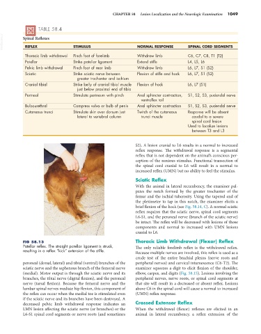
            FIG 58.13                                            Thoracic Limb Withdrawal (Flexor) Reflex
            Patellar reflex. The straight patellar ligament is struck,   The only reliable forelimb reflex is the withdrawal reflex.
            resulting in a reflex “kick” extension of the stifle.   Because multiple nerves are involved, this reflex is used as a
                                                                 crude test of the entire brachial plexus (nerve roots and
            peroneal (dorsal, lateral) and tibial (ventral) branches of the   peripheral nerves) and cervical intumescence (C6-T2). The
            sciatic nerve and the saphenous branch of the femoral nerve   examiner squeezes a digit to elicit flexion of the shoulder,
            (medial). Motor output is through the sciatic nerve and its   elbow, carpus, and digits (Fig. 58.15). Lesions involving the
            branches, the tibial nerve (digital flexion), and the peroneal   peripheral nerves, nerve roots, or spinal cord segments at
            nerve (tarsal flexion). Because the femoral nerve and the   that site will result in a decreased or absent reflex. Lesions
            lumbar spinal nerves mediate hip flexion, this component of   above C6 in the spinal cord will cause a normal to increased
            the reflex can occur when the medial toe is stimulated even   (UMN) reflex response.
            if the sciatic nerve and its branches have been destroyed. A
            decreased pelvic limb withdrawal response indicates an   Crossed Extensor Reflex
            LMN lesion affecting the sciatic nerve (or branches) or the   When the withdrawal (flexor) reflexes are elicited in an
            L6-S1 spinal cord segments or nerve roots (and sometimes   animal in lateral recumbency, a reflex extension of the

