
Page 1080 - Small Animal Internal Medicine, 6th Edition
P. 1080
1052 PART IX Nervous System and Neuromuscular Disorders
cord. If minor stimulation in a paralyzed animal does not cord segments), whereas a behavioral response requires that
elicit a behavioral response such as turning the head, vocal- the sensory spinal cord tracts ascending to the brain also be
VetBooks.ir izing, or trying to bite, then the animal’s ability to perceive a intact.
When LMN paralysis of a limb is evident, mapping the
more severe noxious stimulus such as a hemostat applied to
the nail base (deep pain) should be tested. The spinal tracts
lesion localization to specific peripheral nerves, dorsal nerve
that carry deep pain sensation are small, bilateral, and mul- boundaries of normal and diminished sensation can aid in
tisynaptic and located deep in the spinal cord white matter, roots, or spinal cord segments. The skin should be pinched
so only a very severe bilateral spinal cord lesion will com- with a hemostat and regions of local anesthesia or decreased
pletely disrupt these tracts, making the ability to perceive sensation identified (Fig. 58.19). These results can be com-
deep pain an important prognostic indicator in animals with pared with established maps of cutaneous regions deriving
severe compressive T3-L3 spinal cord injury (Fig. 58.18). It sensory innervation from individual nerves (dermatomes),
is important to remember that withdrawal of the limb indi- allowing the LMN neurologic defect to be precisely localized
cates only an intact reflex arc (peripheral nerve and spinal (see Chapters 65 and 66).
A B
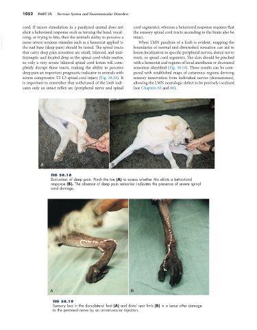
FIG 58.18
Evaluation of deep pain. Pinch the toe (A) to assess whether this elicits a behavioral
response (B). The absence of deep pain sensation indicates the presence of severe spinal
cord damage.
A B
FIG 58.19
Sensory loss in the dorsolateral foot (A) and distal rear limb (B) in a lemur after damage
to the peroneal nerve by an intramuscular injection.