
Page 1079 - Small Animal Internal Medicine, 6th Edition
P. 1079
CHAPTER 58 Lesion Localization and the Neurologic Examination 1051
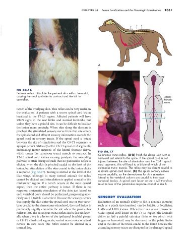
VetBooks.ir C8-T1
A
FIG 58.16
Perineal reflex. Stimulate the perineal skin with a hemostat,
causing the anal sphincter to contract and the tail to C8-T1 Lateral
ventroflex. thoracic
nerve
twitch of the overlying skin. This reflex can be very useful in
the evaluation of patients with a severe spinal cord lesion a
localized to the T3-L3 region. Affected patients will have b
UMN signs in the rear limbs and normal forelimbs, but
unless they have a painful site, it can be difficult to localize
the lesion more precisely. When skin along the dorsum is
pinched, the stimulated sensory nerve from that site enters
the spinal cord and afferent sensory information ascends the
spinal cord in sensory tracts. If the spinal cord is intact
between the site of stimulation and the C8-T1 segments, a B
synapse occurs bilaterally at the C8-T1 spinal cord segments, C
stimulating motor neurons of the lateral thoracic nerve, FIG 58.17
which causes the cutaneous trunci muscle to contract. In Cutaneous trunci reflex. (A-B) Pinch the dorsal skin with a
T3-L3 spinal cord lesions causing paralysis, the ascending hemostat just lateral to the spine. If the spinal cord is not
pathway is often disrupted such that no panniculus reflex is injured between the site of stimulation and the C8-T1 spinal
elicited when the skin is pinched caudal to the level of the cord segments, this will lead to a bilateral twitch of the
lesion, but stimulation of the skin cranial to the lesion elicits cutaneous trunci muscle. The reflex may be absent caudal to
a response (Fig. 58.17). Testing is started at the level of the a severe spinal cord lesion. (C) The spinal sensory nerves
iliac wings, although in many normal animals the reflex course caudally, so the dermatomes for skin sensation
lateral to the vertebral column are caudal to their own
cannot be elicited until stimulation is applied cranial to the vertebral bodies. A spinal cord lesion at site a will therefore
midlumbar region. If a twitch occurs at the most caudal result in loss of the panniculus response caudal to site b.
aspect, then the entire pathway is intact. If there is no
response, systematic stimulation of the skin just lateral to
each vertebral body should be performed, progressing ante-
riorly until a twitch is observed. Because the sensory nerves SENSORY EVALUATION
that supply the skin enter the spinal cord one or two verte- Evaluation of an animal’s ability to feel a noxious stimulus
brae cranial to the dermatome stimulated, the cord lesion is such as a pinch (nociception) can be helpful in localizing
predictably slightly cranial to the site where the panniculus UMN and LMN lesions. When there is a severe transverse
reflex is lost. The cutaneous trunci reflex can be lost unilater- UMN spinal cord lesion in the T3-L3 region, the animal’s
ally when there is a lesion of the ipsilateral brachial plexus ability to feel a painful stimulus (skin or toe pinch with
or C8-T1 spinal cord segments, ventral nerve roots, or spinal fingers or hemostat) may be decreased in the pelvic limbs
nerves. In rare cases this reflex cannot be elicited in a and in the skin of the trunk caudal to the lesion because the
normal dog. ascending sensory tracts are disrupted in the damaged spinal