
Page 1082 - Small Animal Internal Medicine, 6th Edition
P. 1082
1054 PART IX Nervous System and Neuromuscular Disorders
VetBooks.ir
E
G
F
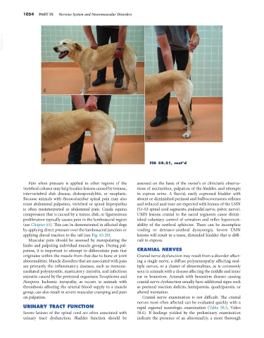
FIG 58.21, cont’d
Pain when pressure is applied to other regions of the assessed on the basis of the owner’s or clinician’s observa-
vertebral column may help localize lesions caused by trauma, tions of micturition, palpation of the bladder, and attempts
intervertebral disk disease, diskospondylitis, or neoplasia. to express urine. A flaccid, easily expressed bladder with
Because animals with thoracolumbar spinal pain may also absent or diminished perineal and bulbocavernosus reflexes
resist abdominal palpation, vertebral or spinal hyperpathia and reduced anal tone are expected with lesions of the LMN
is often misinterpreted as abdominal pain. Cauda equina (S1-S3 spinal cord segments, pudendal nerve, pelvic nerve).
compression that is caused by a tumor, disk, or ligamentous UMN lesions cranial to the sacral segments cause dimin-
proliferation typically causes pain in the lumbosacral region ished voluntary control of urination and reflex hyperexcit-
(see Chapter 65). This can be demonstrated in affected dogs ability of the urethral sphincter. There can be incomplete
by applying direct pressure over the lumbosacral junction or voiding or detrusor-urethral dyssynergia. Severe UMN
applying dorsal traction to the tail (see Fig. 65.20). lesions will result in a tense, distended bladder that is diffi-
Muscular pain should be assessed by manipulating the cult to express.
limbs and palpating individual muscle groups. During pal-
pation, it is important to attempt to differentiate pain that CRANIAL NERVES
originates within the muscle from that due to bone or joint Cranial nerve dysfunction may result from a disorder affect-
abnormalities. Muscle disorders that are associated with pain ing a single nerve, a diffuse polyneuropathy affecting mul-
are primarily the inflammatory diseases, such as immune- tiple nerves, or a cluster of abnormalities, as is commonly
mediated polymyositis, masticatory myositis, and infectious seen in animals with a disease affecting the middle and inner
myositis caused by the protozoal organisms Toxoplasma and ear or brainstem. Animals with brainstem disease causing
Neospora. Ischemic myopathy, as occurs in animals with cranial nerve dysfunction usually have additional signs such
thrombosis affecting the arterial blood supply to a muscle as postural reaction deficits, hemiparesis, quadriparesis, or
group, can also result in severe muscular cramping and pain altered mentation.
on palpation. Cranial nerve examination is not difficult. The cranial
nerves most often affected can be evaluated quickly with a
URINARY TRACT FUNCTION rapid regional neurologic examination (Table 58.5, Video
Severe lesions of the spinal cord are often associated with 58.6). If findings yielded by the preliminary examination
urinary tract dysfunction. Bladder function should be indicate the presence of an abnormality, a more thorough